1F93 | pdb_00001f93

CRYSTAL STRUCTURE OF A COMPLEX BETWEEN THE DIMERIZATION DOMAIN OF HNF-1 ALPHA AND THE COACTIVATOR DCOH


Experimental Data Snapshot

  • Method: X-RAY DIFFRACTION
  • Resolution: 2.60 Å
  • R-Value Free: 
    0.299 (Depositor), 0.296 (DCC) 
  • R-Value Work: 
    0.257 (Depositor), 0.256 (DCC) 
  • R-Value Observed: 
    0.257 (Depositor) 

wwPDB Validation 3D Report Full Report

Validation slider image for 1F93

This is version 1.3 of the entry. See complete history

Literature

Structural basis of dimerization, coactivator recognition and MODY3 mutations in HNF-1alpha.

Rose, R.B.Bayle, J.H.Endrizzi, J.A.Cronk, J.D.Crabtree, G.R.Alber, T.

(2000) Nat Struct Biol 7: 744-748

  • DOI: https://doi.org/10.1038/78966
  • Primary Citation Related Structures: 
    1F93

  • PubMed Abstract: 

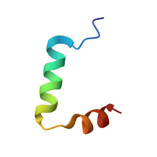
    Maturity-onset diabetes of the young type 3 (MODY3) results from mutations in the transcriptional activator hepatocyte nuclear factor-1alpha (HNF-1alpha). Several MODY3 mutations target the HNF-1alpha dimerization domain (HNF-p1), which binds the coactivator, dimerization cofactor of HNF-1 (DCoH). To define the mechanism of coactivator recognition and the basis for the MODY3 phenotype, we determined the cocrystal structure of the DCoH-HNF-p1 complex and characterized biochemically the effects of MODY3 mutations in HNF-p1. The DCoH-HNF-p1 complex comprises a dimer of dimers in which HNF-p1 forms a unique four-helix bundle. Through rearrangements of interfacial side chains, a single, bifunctional interface in the DCoH dimer mediates both HNF-1alpha binding and formation of a competing, transcriptionally inactive DCoH homotetramer. Consistent with the structure, MODY3 mutations in HNF-p1 reduce activator function by two distinct mechanisms.


  • Organizational Affiliation
    • Department of Molecular and Cell Biology, University of California, Berkeley, California 94720-3206, USA.

Macromolecule Content 

  • Total Structure Weight: 62.18 kDa 
  • Atom Count: 4,106 
  • Modeled Residue Count: 512 
  • Deposited Residue Count: 544 
  • Unique protein chains: 2

Macromolecules

Find similar proteins by:|  3D Structure
Entity ID: 1
MoleculeChains  Sequence LengthOrganismDetailsImage
DIMERIZATION COFACTOR OF HEPATOCYTE NUCLEAR FACTOR 1-ALPHA
A, B, C, D
104Rattus norvegicusMutation(s): 3 
EC: 4.2.1.96
UniProt
Find proteins for P61459 (Rattus norvegicus)
Explore P61459 
Go to UniProtKB:  P61459
Entity Groups
Sequence Clusters30% Identity50% Identity70% Identity90% Identity95% Identity100% Identity
UniProt GroupP61459
Sequence Annotations
Expand
Reference Sequence
Find similar proteins by:|  3D Structure
Entity ID: 2
MoleculeChains  Sequence LengthOrganismDetailsImage
HEPATOCYTE NUCLEAR FACTOR 1-ALPHA
E, F, G, H
32N/AMutation(s): 0 
UniProt
Find proteins for P22361 (Mus musculus)
Explore P22361 
Go to UniProtKB:  P22361
Entity Groups
Sequence Clusters30% Identity50% Identity70% Identity90% Identity95% Identity100% Identity
UniProt GroupP22361
Sequence Annotations
Expand
Reference Sequence

Small Molecules

Modified Residues  1 Unique
IDChains TypeFormula2D DiagramParent
MSE
Query on MSE
A, B, C, D
L-PEPTIDE LINKINGC5 H11 N O2 SeMET

Experimental Data & Validation

Experimental Data

  • Method: X-RAY DIFFRACTION
  • Resolution: 2.60 Å
  • R-Value Free:  0.299 (Depositor), 0.296 (DCC) 
  • R-Value Work:  0.257 (Depositor), 0.256 (DCC) 
  • R-Value Observed: 0.257 (Depositor) 
Space Group: P 1 21 1
Unit Cell:
Length ( Å )Angle ( ˚ )
a = 49.48α = 90
b = 82.75β = 97.83
c = 70.64γ = 90
Software Package:
Software NamePurpose
SHARPphasing
CNSrefinement
MOSFLMdata reduction
CCP4data scaling

Structure Validation

View Full Validation Report


Entry History 

Deposition Data

Revision History  (Full details and data files)

  • Version 1.0: 2000-09-20
    Type: Initial release
  • Version 1.1: 2008-04-27
    Changes: Version format compliance
  • Version 1.2: 2011-07-13
    Changes: Version format compliance
  • Version 1.3: 2024-10-30
    Changes: Data collection, Database references, Derived calculations, Structure summary