1F4W | pdb_00001f4w

CRYSTAL STRUCTURE OF AN ANTI-CARBOHYDRATE ANTIBODY DIRECTED AGAINST VIBRIO CHOLERAE O1 IN COMPLEX WITH ANTIGEN


Experimental Data Snapshot

  • Method: X-RAY DIFFRACTION
  • Resolution: 2.30 Å
  • R-Value Free: 
    0.261 (Depositor) 
  • R-Value Work: 
    0.197 (Depositor) 

Starting Model: experimental
View more details

wwPDB Validation 3D Report Full Report

Validation slider image for 1F4W

This is version 1.5 of the entry. See complete history

Literature

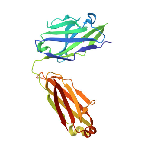
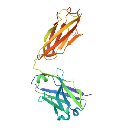
Crystal structure of an anti-carbohydrate antibody directed against Vibrio cholerae O1 in complex with antigen: molecular basis for serotype specificity.

Villeneuve, S.Souchon, H.Riottot, M.M.Mazie, J.C.Lei, P.Glaudemans, C.P.Kovac, P.Fournier, J.M.Alzari, P.M.

(2000) Proc Natl Acad Sci U S A 97: 8433-8438

  • DOI: https://doi.org/10.1073/pnas.060022997
  • Primary Citation Related Structures: 
    1F4W, 1F4X, 1F4Y

  • PubMed Abstract: 

    The crystal structure of the murine Fab S-20-4 from a protective anti-cholera Ab specific for the lipopolysaccharide Ag of the Ogawa serotype has been determined in its unliganded form and in complex with synthetic fragments of the Ogawa O-specific polysaccharide (O-SP). The upstream terminal O-SP monosaccharide is shown to be the primary antigenic determinant. Additional perosamine residues protrude outwards from the Ab surface and contribute only marginally to the binding affinity and specificity. A complementary water-excluding hydrophobic interface and five Ab-Ag hydrogen bonds are crucial for carbohydrate recognition. The structure reported here explains the serotype specificity of anti-Ogawa Abs and provides a rational basis toward the development of a synthetic carbohydrate-based anti-cholera vaccine.


  • Organizational Affiliation
    • Unité de Biochimie Structurale (Centre National de la Recherche Scientifique, Unité de Recherche Associée 2185), Laboratoire d'Ingénierie des Anticorps, Institut Pasteur, Paris, France.

Macromolecule Content 

  • Total Structure Weight: 45.79 kDa 
  • Atom Count: 3,205 
  • Modeled Residue Count: 426 
  • Deposited Residue Count: 426 
  • Unique protein chains: 2

Macromolecules

Find similar proteins by:|  3D Structure
Entity ID: 1
MoleculeChains  Sequence LengthOrganismDetailsImage
ANTIBODY S-20-4, FAB FRAGMENT, LIGHT CHAINA [auth L]210Mus musculusMutation(s): 0 
Find similar proteins by:|  3D Structure
Entity ID: 2
MoleculeChains  Sequence LengthOrganismDetailsImage
ANTIBODY S-20-4, FAB FRAGMENT, HEAVY CHAINB [auth H]216Mus musculusMutation(s): 0 

Experimental Data & Validation

Experimental Data

  • Method: X-RAY DIFFRACTION
  • Resolution: 2.30 Å
  • R-Value Free:  0.261 (Depositor) 
  • R-Value Work:  0.197 (Depositor) 
Space Group: P 1 21 1
Unit Cell:
Length ( Å )Angle ( ˚ )
a = 45.67α = 90
b = 113.32β = 100.69
c = 46.21γ = 90
Software Package:
Software NamePurpose
DENZOdata reduction
SCALEPACKdata scaling
AMoREphasing
REFMACrefinement

Structure Validation

View Full Validation Report


Entry History 

Deposition Data

Revision History  (Full details and data files)

  • Version 1.0: 2000-08-02
    Type: Initial release
  • Version 1.1: 2008-04-27
    Changes: Version format compliance
  • Version 1.2: 2011-07-13
    Changes: Version format compliance
  • Version 1.3: 2018-02-07
    Changes: Experimental preparation
  • Version 1.4: 2024-04-03
    Changes: Data collection, Database references, Refinement description
  • Version 1.5: 2024-10-30
    Changes: Structure summary