1F4N | pdb_00001f4n

C2 CRYSTAL STRUCTURE OF ALA2ILE2-6, A VERSION OF ROP WITH A REPACKED HYDROPHOBIC CORE AND A NEW FOLD.


Experimental Data Snapshot

  • Method: X-RAY DIFFRACTION
  • Resolution: 1.90 Å
  • R-Value Free: 
    0.237 (Depositor), 0.231 (DCC) 
  • R-Value Work: 
    0.204 (Depositor), 0.195 (DCC) 

wwPDB Validation 3D Report Full Report

Validation slider image for 1F4N

This is version 1.4 of the entry. See complete history

Literature

Dramatic structural and thermodynamic consequences of repacking a protein's hydrophobic core.

Willis, M.A.Bishop, B.Regan, L.Brunger, A.T.

(2000) Structure 8: 1319-1328

  • DOI: https://doi.org/10.1016/s0969-2126(00)00544-x
  • Primary Citation Related Structures: 
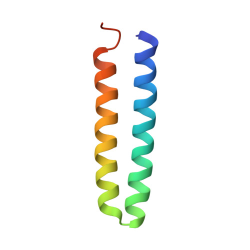
    1F4M, 1F4N

  • PubMed Abstract: 

    Rop is an RNA binding, dimeric, four-helix bundle protein with a well-defined, regular hydrophobic core ideally suited for redesign studies. A family of Rop variants in which the hydrophobic core was systematically redesigned has previously been created and characterized. We present a structural and thermodynamic analysis of Ala2Ile2-6, a variant of Rop with an extensively redesigned hydrophobic core. The structure of Ala2Ile2-6 reveals a completely new fold formed by a conformational "flip" of the two protomers around the dimeric interface. The free-energy profile of Ala2Ile2-6 is also very different from that of wild-type Rop. Ala2Ile2-6 has a higher melting temperature than Rop, but undergoes a slightly smaller free-energy change on unfolding. The structure of Ala2Ile2-6, along with molecular modeling results, demonstrate the importance of tight packing of core residues and the adoption of favorable core side chain rotamer values in determining helix-helix interactions in the four-helix bundle fold. Structural disorder at the N and C termini of Ala2Ile2-6 provides a basis for the large differences in the enthalpy and entropy of Ala2Ile2-6 folding compared with wildtype Rop.


  • Organizational Affiliation
    • Department of Molecular Biophysics and Biochemistry, Yale University, New Haven, Connecticut 06520, USA.

Macromolecule Content 

  • Total Structure Weight: 14.39 kDa 
  • Atom Count: 965 
  • Modeled Residue Count: 111 
  • Deposited Residue Count: 126 
  • Unique protein chains: 1

Macromolecules

Find similar proteins by:|  3D Structure
Entity ID: 1
MoleculeChains  Sequence LengthOrganismDetailsImage
ROP ALA2ILE2-6
A, B
63Escherichia coliMutation(s): 10 
UniProt
Find proteins for P03051 (Escherichia coli)
Explore P03051 
Go to UniProtKB:  P03051
Entity Groups
Sequence Clusters30% Identity50% Identity70% Identity90% Identity95% Identity100% Identity
UniProt GroupP03051
Sequence Annotations
Expand
Reference Sequence

Experimental Data & Validation

Experimental Data

  • Method: X-RAY DIFFRACTION
  • Resolution: 1.90 Å
  • R-Value Free:  0.237 (Depositor), 0.231 (DCC) 
  • R-Value Work:  0.204 (Depositor), 0.195 (DCC) 
Space Group: C 1 2 1
Unit Cell:
Length ( Å )Angle ( ˚ )
a = 82.617α = 90
b = 36.488β = 121.205
c = 47.727γ = 90
Software Package:
Software NamePurpose
CNSrefinement
DENZOdata reduction
SCALEPACKdata scaling
CNSphasing

Structure Validation

View Full Validation Report


Entry History 

Deposition Data

Revision History  (Full details and data files)

  • Version 1.0: 2001-01-10
    Type: Initial release
  • Version 1.1: 2008-04-27
    Changes: Version format compliance
  • Version 1.2: 2011-07-13
    Changes: Version format compliance
  • Version 1.3: 2021-11-03
    Changes: Database references, Derived calculations
  • Version 1.4: 2024-02-07
    Changes: Data collection