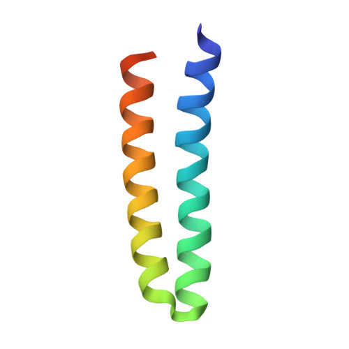
1F4M

P3(2) CRYSTAL STRUCTURE OF ALA2ILE2-6, A VERSION OF ROP WITH A REPACKED HYDROPHOBIC CORE AND A NEW FOLD.


Experimental Data Snapshot

  • Method: X-RAY DIFFRACTION
  • Resolution: 2.25 Å
  • R-Value Free: 0.282 
  • R-Value Work: 0.233 

wwPDB Validation   3D Report Full Report


This is version 1.4 of the entry. See complete history


Literature

Dramatic structural and thermodynamic consequences of repacking a protein's hydrophobic core.

Willis, M.A.Bishop, B.Regan, L.Brunger, A.T.

(2000) Structure 8: 1319-1328

  • DOI: https://doi.org/10.1016/s0969-2126(00)00544-x
  • Primary Citation of Related Structures:  
    1F4M, 1F4N

  • PubMed Abstract: 

    Rop is an RNA binding, dimeric, four-helix bundle protein with a well-defined, regular hydrophobic core ideally suited for redesign studies. A family of Rop variants in which the hydrophobic core was systematically redesigned has previously been created and characterized. We present a structural and thermodynamic analysis of Ala2Ile2-6, a variant of Rop with an extensively redesigned hydrophobic core. The structure of Ala2Ile2-6 reveals a completely new fold formed by a conformational "flip" of the two protomers around the dimeric interface. The free-energy profile of Ala2Ile2-6 is also very different from that of wild-type Rop. Ala2Ile2-6 has a higher melting temperature than Rop, but undergoes a slightly smaller free-energy change on unfolding. The structure of Ala2Ile2-6, along with molecular modeling results, demonstrate the importance of tight packing of core residues and the adoption of favorable core side chain rotamer values in determining helix-helix interactions in the four-helix bundle fold. Structural disorder at the N and C termini of Ala2Ile2-6 provides a basis for the large differences in the enthalpy and entropy of Ala2Ile2-6 folding compared with wildtype Rop.


  • Organizational Affiliation

    Department of Molecular Biophysics and Biochemistry, Yale University, New Haven, Connecticut 06520, USA.


Macromolecules
Find similar proteins by:  (by identity cutoff)  |  3D Structure
Entity ID: 1
MoleculeChains Sequence LengthOrganismDetailsImage
ROP ALA2ILE2-6
A, B, C, D, E
A, B, C, D, E, F
63Escherichia coliMutation(s): 10 
UniProt
Find proteins for P03051 (Escherichia coli)
Explore P03051 
Go to UniProtKB:  P03051
Entity Groups  
Sequence Clusters30% Identity50% Identity70% Identity90% Identity95% Identity100% Identity
UniProt GroupP03051
Sequence Annotations
Expand
  • Reference Sequence
Experimental Data & Validation

Experimental Data

  • Method: X-RAY DIFFRACTION
  • Resolution: 2.25 Å
  • R-Value Free: 0.282 
  • R-Value Work: 0.233 
  • Space Group: P 32
Unit Cell:
Length ( Å )Angle ( ˚ )
a = 73.092α = 90
b = 73.092β = 90
c = 65.921γ = 120
Software Package:
Software NamePurpose
CNSrefinement
DENZOdata reduction
SCALEPACKdata scaling
CNSphasing

Structure Validation

View Full Validation Report


Entry History 

Deposition Data

Revision History  (Full details and data files)

  • Version 1.0: 2001-01-10
    Type: Initial release
  • Version 1.1: 2008-04-27
    Changes: Version format compliance
  • Version 1.2: 2011-07-13
    Changes: Version format compliance
  • Version 1.3: 2021-11-03
    Changes: Database references, Derived calculations
  • Version 1.4: 2024-02-07
    Changes: Data collection