1F36 | pdb_00001f36

THE CRYSTAL STRUCTURE OF FIS MUTANT K36E REVEALS THAT THE TRANSACTIVATION REGION OF THE FIS PROTEIN CONTAINS EXTENDED MOBILE BETA-HAIRPIN ARMS


Experimental Data Snapshot

  • Method: X-RAY DIFFRACTION
  • Resolution: 2.65 Å
  • R-Value Free: 
    0.277 (Depositor), 0.274 (DCC) 
  • R-Value Work: 
    0.222 (Depositor), 0.212 (DCC) 
  • R-Value Observed: 
    0.222 (Depositor) 

Starting Model: experimental
View more details

wwPDB Validation 3D Report Full Report

Validation slider image for 1F36

This is version 1.5 of the entry. See complete history

Literature

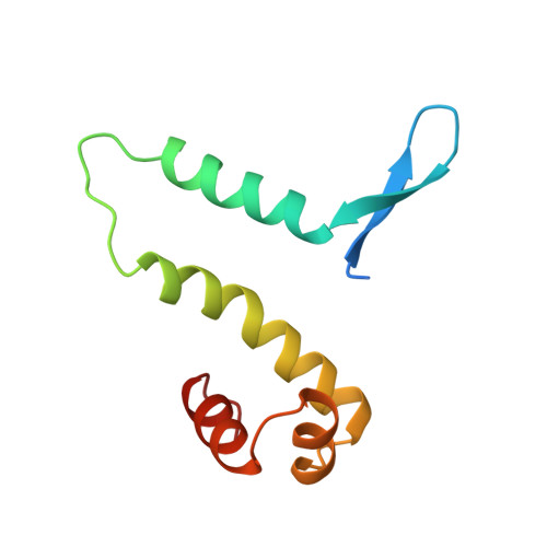
The transactivation region of the fis protein that controls site-specific DNA inversion contains extended mobile beta-hairpin arms.

Safo, M.K.Yang, W.Z.Corselli, L.Cramton, S.E.Yuan, H.S.Johnson, R.C.

(1997) EMBO J 16: 6860-6873

  • DOI: https://doi.org/10.1093/emboj/16.22.6860
  • Primary Citation Related Structures: 
    1F36

  • PubMed Abstract: 

    The Fis protein regulates site-specific DNA inversion catalyzed by a family of DNA invertases when bound to a cis-acting recombinational enhancer. As is often found for transactivation domains, previous crystal structures have failed to resolve the conformation of the N-terminal inversion activation region within the Fis dimer. A new crystal form of a mutant Fis protein now reveals that the activation region contains two beta-hairpin arms that protrude over 20 A from the protein core. Saturation mutagenesis identified the regulatory and structurally important amino acids. The most critical activating residues are located near the tips of the beta-arms. Disulfide cross-linking between the beta-arms demonstrated that they are highly flexible in solution and that efficient inversion activation can occur when the beta-arms are covalently linked together. The emerging picture for this regulatory motif is that contacts with the recombinase at the tip of the mobile beta-arms activate the DNA invertase in the context of an invertasome complex.


  • Organizational Affiliation
    • Institute of Molecular Biology, Academia Sinica, Taipei, Taiwan 11529, Republic of China.

Macromolecule Content 

  • Total Structure Weight: 22.51 kDa 
  • Atom Count: 1,438 
  • Modeled Residue Count: 178 
  • Deposited Residue Count: 196 
  • Unique protein chains: 1

Macromolecules

Find similar proteins by:|  3D Structure
Entity ID: 1
MoleculeChains  Sequence LengthOrganismDetailsImage
FIS
A, B
98Escherichia coliMutation(s): 1 
Gene Names: FIS
UniProt
Find proteins for P0A6R3 (Escherichia coli (strain K12))
Explore P0A6R3 
Go to UniProtKB:  P0A6R3
Entity Groups
Sequence Clusters30% Identity50% Identity70% Identity90% Identity95% Identity100% Identity
UniProt GroupP0A6R3
Sequence Annotations
Expand
Reference Sequence

Experimental Data & Validation

Experimental Data

  • Method: X-RAY DIFFRACTION
  • Resolution: 2.65 Å
  • R-Value Free:  0.277 (Depositor), 0.274 (DCC) 
  • R-Value Work:  0.222 (Depositor), 0.212 (DCC) 
  • R-Value Observed: 0.222 (Depositor) 
Space Group: P 65 2 2
Unit Cell:
Length ( Å )Angle ( ˚ )
a = 51.9α = 90
b = 51.9β = 90
c = 294.93γ = 120
Software Package:
Software NamePurpose
AMoREphasing
X-PLORrefinement
MSCdata reduction
MSCdata scaling

Structure Validation

View Full Validation Report


Entry History 

Deposition Data

Revision History  (Full details and data files)

  • Version 1.0: 1997-12-24
    Type: Initial release
  • Version 1.1: 2008-03-24
    Changes: Version format compliance
  • Version 1.2: 2011-07-13
    Changes: Version format compliance
  • Version 1.3: 2021-11-03
    Changes: Database references
  • Version 1.4: 2024-02-07
    Changes: Data collection
  • Version 1.5: 2024-04-03
    Changes: Refinement description