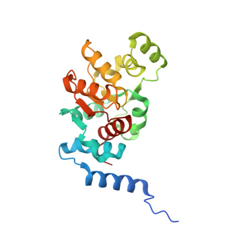
1F2V

CRYSTAL STRUCTURE ANALYSIS OF PRECORRIN-8X METHYLMUTASE OF AEROBIC VITAMIN B12 SYNTHESIS


Experimental Data Snapshot

  • Method: X-RAY DIFFRACTION
  • Resolution: 2.10 Å
  • R-Value Free: 0.236 
  • R-Value Work: 0.183 
  • R-Value Observed: 0.187 

wwPDB Validation   3D Report Full Report


This is version 1.3 of the entry. See complete history


Literature

Crystal structure of precorrin-8x methyl mutase.

Shipman, L.W.Li, D.Roessner, C.A.Scott, A.I.Sacchettini, J.C.

(2001) Structure 9: 587-596

  • DOI: https://doi.org/10.1016/s0969-2126(01)00618-9
  • Primary Citation of Related Structures:  
    1F2V, 1I1H

  • PubMed Abstract: 

    The crystal structure of precorrin-8x methyl mutase (CobH), an enzyme of the aerobic pathway to vitamin B12, provides evidence that the mechanism for methyl migration can plausibly be regarded as an allowed [1,5]-sigmatropic shift of a methyl group from C-11 to C-12 at the C ring of precorrin-8x to afford hydrogenobyrinic acid. The dimeric structure of CobH creates a set of shared active sites that readily discriminate between different tautomers of precorrin-8x and select a discrete tautomer for sigmatropic rearrangement. The active site contains a strictly conserved histidine residue close to the site of methyl migration in ring C of the substrate. Analysis of the structure with bound product suggests that the [1,5]-sigmatropic shift proceeds by protonation of the ring C nitrogen, leading to subsequent methyl migration.


  • Organizational Affiliation

    Department of Chemistry, Texas A&M University, College Station, TX 77843, USA. sacchett@tamu.edu


Macromolecules
Find similar proteins by:  (by identity cutoff)  |  3D Structure
Entity ID: 1
MoleculeChains Sequence LengthOrganismDetailsImage
PRECORRIN-8X METHYLMUTASE219Pseudomonas denitrificans (nom. rej.)Mutation(s): 0 
EC: 5.4.1.2 (PDB Primary Data), 5.4.99.61 (UniProt)
UniProt
Find proteins for P21638 (Sinorhizobium sp)
Explore P21638 
Go to UniProtKB:  P21638
Entity Groups  
Sequence Clusters30% Identity50% Identity70% Identity90% Identity95% Identity100% Identity
UniProt GroupP21638
Sequence Annotations
Expand
  • Reference Sequence
Experimental Data & Validation

Experimental Data

  • Method: X-RAY DIFFRACTION
  • Resolution: 2.10 Å
  • R-Value Free: 0.236 
  • R-Value Work: 0.183 
  • R-Value Observed: 0.187 
  • Space Group: C 1 2 1
Unit Cell:
Length ( Å )Angle ( ˚ )
a = 90.1α = 90
b = 36.6β = 113.8
c = 60.3γ = 90
Software Package:
Software NamePurpose
SOLVEphasing
CNSrefinement
DENZOdata reduction
SCALEPACKdata scaling

Structure Validation

View Full Validation Report


Entry History 

Deposition Data

Revision History  (Full details and data files)

  • Version 1.0: 2001-07-18
    Type: Initial release
  • Version 1.1: 2008-04-27
    Changes: Version format compliance
  • Version 1.2: 2011-07-13
    Changes: Derived calculations, Version format compliance
  • Version 1.3: 2024-02-07
    Changes: Data collection, Database references