1EZJ | pdb_00001ezj

CRYSTAL STRUCTURE OF THE MULTIMERIZATION DOMAIN OF THE PHOSPHOPROTEIN FROM SENDAI VIRUS


Experimental Data Snapshot

  • Method: X-RAY DIFFRACTION
  • Resolution: 1.90 Å
  • R-Value Free: 
    0.293 (Depositor), 0.298 (DCC) 
  • R-Value Work: 
    0.249 (Depositor), 0.253 (DCC) 
  • R-Value Observed: 
    0.255 (Depositor) 

wwPDB Validation 3D Report Full Report

Validation slider image for 1EZJ

This is version 1.4 of the entry. See complete history

Literature

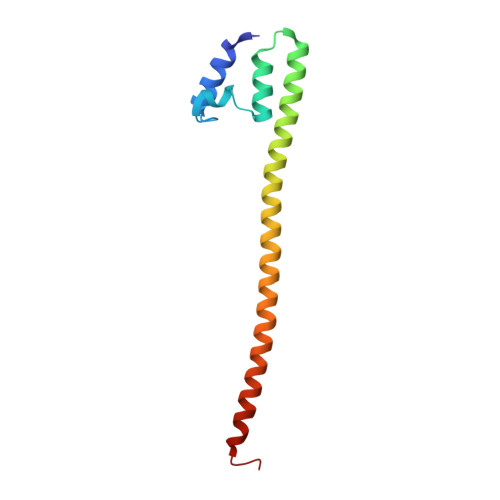
Tetrameric coiled coil domain of Sendai virus phosphoprotein.

Tarbouriech, N.Curran, J.Ruigrok, R.W.Burmeister, W.P.

(2000) Nat Struct Biol 7: 777-781

  • DOI: https://doi.org/10.1038/79013
  • Primary Citation Related Structures: 
    1EZJ

  • PubMed Abstract: 

    The high resolution X-ray structure of the Sendai virus oligomerization domain reveals a homotetrameric coiled coil structure with many details that are different from classic coiled coils with canonical hydrophobic heptad repeats. Alternatives to the classic knobs-into-holes packing lead to differences in supercoil pitch and diameter that allow water molecules inside the core. This open and more hydrophilic structure does not seem to be destabilized by mutations that would be expected to disrupt classic coiled coils.


  • Organizational Affiliation
    • European Synchrotron Radiation Facility, Grenoble cedex, France.

Macromolecule Content 

  • Total Structure Weight: 13.39 kDa 
  • Atom Count: 1,064 
  • Modeled Residue Count: 114 
  • Deposited Residue Count: 115 
  • Unique protein chains: 1

Macromolecules

Find similar proteins by:|  3D Structure
Entity ID: 1
MoleculeChains  Sequence LengthOrganismDetailsImage
NUCLEOCAPSID PHOSPHOPROTEIN115Sendai virus (strain Harris)Mutation(s): 0 
EC: 2.7.7.48
UniProt
Find proteins for P04859 (Sendai virus (strain Harris))
Explore P04859 
Go to UniProtKB:  P04859
Entity Groups
Sequence Clusters30% Identity50% Identity70% Identity90% Identity95% Identity100% Identity
UniProt GroupP04859
Sequence Annotations
Expand
Reference Sequence

Experimental Data & Validation

Experimental Data

  • Method: X-RAY DIFFRACTION
  • Resolution: 1.90 Å
  • R-Value Free:  0.293 (Depositor), 0.298 (DCC) 
  • R-Value Work:  0.249 (Depositor), 0.253 (DCC) 
  • R-Value Observed: 0.255 (Depositor) 
Space Group: P 4 21 2
Unit Cell:
Length ( Å )Angle ( ˚ )
a = 48.584α = 90
b = 48.584β = 90
c = 100.847γ = 90
Software Package:
Software NamePurpose
SHARPphasing
CNSrefinement
DENZOdata reduction
CCP4data scaling

Structure Validation

View Full Validation Report


Entry History 

Deposition Data

Revision History  (Full details and data files)

  • Version 1.0: 2000-09-20
    Type: Initial release
  • Version 1.1: 2008-04-27
    Changes: Version format compliance
  • Version 1.2: 2011-07-13
    Changes: Derived calculations, Version format compliance
  • Version 1.3: 2011-11-16
    Changes: Atomic model
  • Version 1.4: 2024-02-07
    Changes: Data collection, Database references, Derived calculations