1ETN | pdb_00001etn

MOLECULAR STRUCTURE OF THE TOXIC DOMAIN OF HEAT-STABLE ENTEROTOXIN PRODUCED BY A PATHOGENIC STRAIN OF ESCHERICHIA COLI


Experimental Data Snapshot

  • Method: X-RAY DIFFRACTION
  • Resolution: 0.89 Å
  • R-Value Work: 
    0.088 (Depositor), 0.144 (DCC) 

wwPDB Validation 3D Report Full Report

Validation slider image for 1ETN

This is version 1.5 of the entry. See complete history

Literature

Molecular structure of the toxin domain of heat-stable enterotoxin produced by a pathogenic strain of Escherichia coli. A putative binding site for a binding protein on rat intestinal epithelial cell membranes.

Ozaki, H.Sato, T.Kubota, H.Hata, Y.Katsube, Y.Shimonishi, Y.

(1991) J Biological Chem 266: 5934-5941

  • Primary Citation Related Structures: 
    1ETN

  • PubMed Abstract: 

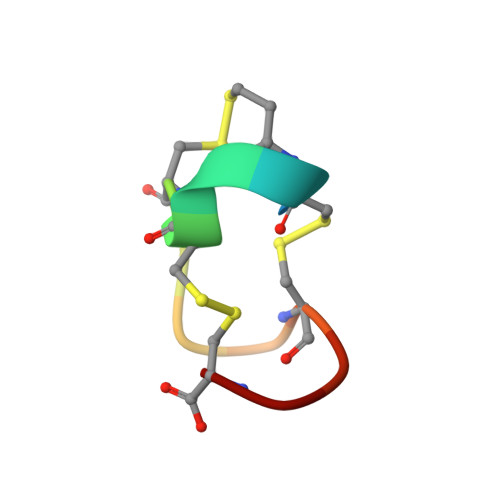
    Heat-stable enterotoxins are a family of toxin peptides that are produced by enterotoxigenic Escherichia coli and consist of 18 and 19 amino acid residues (Aimoto, S., Takao, T., Shimonishi, Y., Hara, S., Takeda, T., Takeda, Y., and Miwatani, T. (1982) Eur. J. Biochem. 129, 257-263). A synthetic fully toxic analog of the enterotoxin, Mpr5-STp(5-17), where Mpr is beta-mercaptopropionic acid and which consists of 13 amino acid residues from Cys5 to Cys17 in a heat-stable enterotoxin but is deaminated at its N terminus (Kubota, H., Hidaka, Y., Ozaki, H., Ito, H., Hirayama, T., Takeda, Y., and Shimonishi, Y. (1989) Biochem. Biophys. Res. Commun. 161, 229-235), has been crystalized from water, and its crystal structure has been solved by a direct method and refined by least square procedures to give an R factor of 0.089. The crystal belongs to the orthorhombic space group P2(1)2(1)2(1) with unit cell constants a = 21.010 (2) A, b = 27.621 (4) A, and c = 12.781 (1) A. The asymmetric unit of the crystals contains one peptide molecule with 13 water molecules. A right-hand spiral peptide backbone extends throughout the molecule. Three beta-turns are located along this spiral and fixed tightly by three intramolecular disulfide linkages. The actual structure predicts the biniding region on the enterotoxin to the receptor protein on the membrane of rat intestinal epithelial cells.


  • Organizational Affiliation
    • Institute for Protein Research, Osaka University, Japan.

Macromolecule Content 

  • Total Structure Weight: 1.26 kDa 
  • Atom Count: 95 
  • Modeled Residue Count: 13 
  • Deposited Residue Count: 13 
  • Unique protein chains: 1

Macromolecules

Find similar proteins by:  Sequence   |   3D Structure  
Entity ID: 1
MoleculeChains  Sequence LengthOrganismDetailsImage
5-BETA-MERCAPTOPROPIONATE HEAT-STABLE ENTEROTOXIN13Escherichia coliMutation(s): 0 
Membrane Entity: Yes 
UniProt
Find proteins for P01559 (Escherichia coli)
Explore P01559 
Go to UniProtKB:  P01559
Entity Groups
Sequence Clusters30% Identity50% Identity70% Identity90% Identity95% Identity100% Identity
UniProt GroupP01559
Sequence Annotations
Expand
Reference Sequence

Experimental Data & Validation

Experimental Data

  • Method: X-RAY DIFFRACTION
  • Resolution: 0.89 Å
  • R-Value Work:  0.088 (Depositor), 0.144 (DCC) 
Space Group: P 21 21 21
Unit Cell:
Length ( Å )Angle ( ˚ )
a = 21.01α = 90
b = 27.621β = 90
c = 12.781γ = 90
Software Package:
Software NamePurpose
FMLS/VPrefinement

Structure Validation

View Full Validation Report


Entry History 

Deposition Data

Revision History  (Full details and data files)

  • Version 1.0: 1996-01-29
    Type: Initial release
  • Version 1.1: 2008-03-03
    Changes: Version format compliance
  • Version 1.2: 2011-07-13
    Changes: Version format compliance
  • Version 1.3: 2013-06-19
    Changes: Other
  • Version 1.4: 2017-11-29
    Changes: Derived calculations, Other
  • Version 1.5: 2025-03-26
    Changes: Data collection, Database references, Derived calculations, Structure summary