1EQ9 | pdb_00001eq9

CRYSTAL STRUCTURE OF FIRE ANT CHYMOTRYPSIN COMPLEXED TO PMSF


Experimental Data Snapshot

  • Method: X-RAY DIFFRACTION
  • Resolution: 1.70 Å
  • R-Value Free: 
    0.242 (Depositor) 
  • R-Value Work: 
    0.209 (Depositor) 
  • R-Value Observed: 
    0.209 (Depositor) 

wwPDB Validation 3D Report Full Report

Validation slider image for 1EQ9

This is version 1.3 of the entry. See complete history

Literature

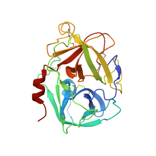
The structure of an insect chymotrypsin.

Botos, I.Meyer, E.Nguyen, M.Swanson, S.M.Koomen, J.M.Russell, D.H.Meyer, E.F.

(2000) J Mol Biology 298: 895-901

  • DOI: https://doi.org/10.1006/jmbi.2000.3699
  • Primary Citation Related Structures: 
    1EQ9

  • PubMed Abstract: 

    The South American imported fire ant (Solenopsis invicta), without natural enemies in the United States, widely infests the southern United States, causing more than a half billion dollars in health and agriculture-related damage annually in Texas alone. Fire ants are resistant to most insecticides, so control will require a more fundamental understanding of their biochemistry and metabolism leading to the design of selective, ecologically safe insecticides. The 4th instar larvae play a crucial role in the nutrition of the colony by secreting proteinases (especially chymotrypsin) which digest food products for the entire colony. The first structure of an ant proteolytic enzyme, fire ant chymotrypsin, was determined to atomic resolution (1.7 A). A structural comparison of the ant and mammalian structures confirms the "universality" of the serine proteinase motif and reveals a difference at residues 147-148, which are proteolytically removed in the bovine enzyme but are firmly intact in the ant chymotrypsin, suggesting a different activation mechanism for the latter. Likewise, the absence of the covalently attached propeptide domain (1-15) further suggests an uncharacteristic activation mechanism. The presence of Gly189 in the S1 site is an atypical feature of this chymotrypsin and is comparable only to human leukocyte elastase, hornet chymotrypsin and fiddler crab collagenase. Binding studies confirm the chymotrypsin nature of this novel enzyme.


  • Organizational Affiliation
    • Department of Biochemistry and Biophysics, Texas A&M University, TX 77843, USA.

Macromolecule Content 

  • Total Structure Weight: 48.42 kDa 
  • Atom Count: 3,925 
  • Modeled Residue Count: 444 
  • Deposited Residue Count: 444 
  • Unique protein chains: 1

Macromolecules

Find similar proteins by:|  3D Structure
Entity ID: 1
MoleculeChains  Sequence LengthOrganismDetailsImage
CHYMOTRYPSIN
A, B
222Solenopsis invictaMutation(s): 0 
EC: 3.4.21.1
UniProt
Find proteins for Q7SIG2 (Solenopsis invicta)
Explore Q7SIG2 
Go to UniProtKB:  Q7SIG2
Entity Groups
Sequence Clusters30% Identity50% Identity70% Identity90% Identity95% Identity100% Identity
UniProt GroupQ7SIG2
Sequence Annotations
Expand
Reference Sequence

Small Molecules

Ligands 1 Unique
IDChains Name / Formula / InChI Key2D Diagram3D Interactions
PMS

Query on PMS


Download:Ideal Coordinates CCD File
C [auth A],
D [auth B]
phenylmethanesulfonic acid
C7 H8 O3 S
NIXKBAZVOQAHGC-UHFFFAOYSA-N

Experimental Data & Validation

Experimental Data

  • Method: X-RAY DIFFRACTION
  • Resolution: 1.70 Å
  • R-Value Free:  0.242 (Depositor) 
  • R-Value Work:  0.209 (Depositor) 
  • R-Value Observed: 0.209 (Depositor) 
Space Group: P 21 21 21
Unit Cell:
Length ( Å )Angle ( ˚ )
a = 61.12α = 90
b = 85.84β = 90
c = 97.61γ = 90
Software Package:
Software NamePurpose
DENZOdata reduction
SCALEPACKdata scaling
AMoREphasing
CNSrefinement

Structure Validation

View Full Validation Report


Entry History 

Deposition Data

Revision History  (Full details and data files)

  • Version 1.0: 2000-10-03
    Type: Initial release
  • Version 1.1: 2008-04-27
    Changes: Version format compliance
  • Version 1.2: 2011-07-13
    Changes: Version format compliance
  • Version 1.3: 2024-10-30
    Changes: Data collection, Database references, Derived calculations, Structure summary