1EPU

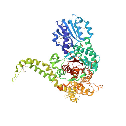
X-RAY crystal structure of neuronal SEC1 from squid


Experimental Data Snapshot

  • Method: X-RAY DIFFRACTION
  • Resolution: 2.40 Å
  • R-Value Free: 0.277 
  • R-Value Work: 0.240 
  • R-Value Observed: 0.251 

wwPDB Validation   3D Report Full Report


This is version 1.5 of the entry. See complete history


Literature

The X-ray crystal structure of neuronal Sec1 from squid sheds new light on the role of this protein in exocytosis.

Bracher, A.Perrakis, A.Dresbach, T.Betz, H.Weissenhorn, W.

(2000) Structure 8: 685-694

  • DOI: https://doi.org/10.1016/s0969-2126(00)00156-8
  • Primary Citation of Related Structures:  
    1EPU

  • PubMed Abstract: 

    Sec1-like molecules have been implicated in a variety of eukaryotic vesicle transport processes including neurotransmitter release by exocytosis. They regulate vesicle transport by binding to a t-SNARE from the syntaxin family. This process is thought to prevent SNARE complex formation, a protein complex required for membrane fusion. Whereas Sec1 molecules are essential for neurotransmitter release and other secretory events, their interaction with syntaxin molecules seems to represent a negative regulatory step in secretion. Here we report the X-ray crystal structure of a neuronal Sec1 homologue from squid, s-Sec1, at 2.4 A resolution. Neuronal s-Sec1 is a modular protein that folds into a V-shaped three-domain assembly. Peptide and mutagenesis studies are discussed with respect to the mechanism of Sec1 regulation. Comparison of the structure of squid s-Sec1 with the previously determined structure of rat neuronal Sec1 (n-Sec1) bound to syntaxin-1a indicates conformational rearrangements in domain III induced by syntaxin binding. The crystal structure of s-Sec1 provides the molecular scaffold for a number of molecular interactions that have been reported to affect Sec1 function. The structural differences observed between s-Sec1 and the structure of a rat n-Sec1-syntaxin-1a complex suggest that local conformational changes are sufficient to release syntaxin-1a from neuronal Sec1, an active process that is thought to involve additional effector molecule(s).


  • Organizational Affiliation

    European Molecular Biology Laboratory (EMBL), Grenoble, 38000, France.


Macromolecules
Find similar proteins by:  (by identity cutoff)  |  3D Structure
Entity ID: 1
MoleculeChains Sequence LengthOrganismDetailsImage
S-SEC1591Doryteuthis pealeiiMutation(s): 0 
UniProt
Find proteins for O62547 (Doryteuthis pealeii)
Explore O62547 
Go to UniProtKB:  O62547
Entity Groups  
Sequence Clusters30% Identity50% Identity70% Identity90% Identity95% Identity100% Identity
UniProt GroupO62547
Sequence Annotations
Expand
  • Reference Sequence
Small Molecules
Modified Residues  1 Unique
IDChains TypeFormula2D DiagramParent
MSE
Query on MSE
A
L-PEPTIDE LINKINGC5 H11 N O2 SeMET
Experimental Data & Validation

Experimental Data

  • Method: X-RAY DIFFRACTION
  • Resolution: 2.40 Å
  • R-Value Free: 0.277 
  • R-Value Work: 0.240 
  • R-Value Observed: 0.251 
  • Space Group: P 1 21 1
Unit Cell:
Length ( Å )Angle ( ˚ )
a = 49.063α = 90
b = 121.989β = 104.53
c = 69.046γ = 90
Software Package:
Software NamePurpose
SnBphasing
SHARPphasing
DMMultimodel building
CNSrefinement
DENZOdata reduction
SCALEPACKdata scaling
DMMultiphasing

Structure Validation

View Full Validation Report


Entry History 

Deposition Data

Revision History  (Full details and data files)

  • Version 1.0: 2000-08-09
    Type: Initial release
  • Version 1.1: 2008-04-27
    Changes: Version format compliance
  • Version 1.2: 2011-07-13
    Changes: Version format compliance
  • Version 1.3: 2011-07-27
    Changes: Atomic model, Data collection
  • Version 1.4: 2017-10-04
    Changes: Refinement description
  • Version 1.5: 2024-10-30
    Changes: Data collection, Database references, Derived calculations, Structure summary