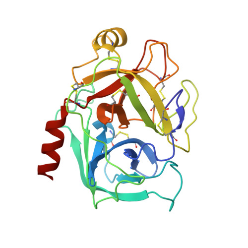
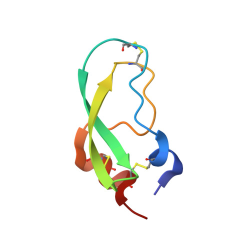
1EJM

CRYSTAL STRUCTURE OF THE BPTI ALA16LEU MUTANT IN COMPLEX WITH BOVINE TRYPSIN


Experimental Data Snapshot

  • Method: X-RAY DIFFRACTION
  • Resolution: 1.85 Å
  • R-Value Free: 0.233 
  • R-Value Work: 0.211 
  • R-Value Observed: 0.212 

wwPDB Validation   3D Report Full Report


This is version 1.7 of the entry. See complete history


Literature

Substitutions at the P(1) position in BPTI strongly affect the association energy with serine proteinases.

Grzesiak, A.Helland, R.Smalas, A.O.Krowarsch, D.Dadlez, M.Otlewski, J.

(2000) J Mol Biol 301: 205-217

  • DOI: https://doi.org/10.1006/jmbi.2000.3935
  • Primary Citation of Related Structures:  
    1EJM

  • PubMed Abstract: 

    The role of the S(1) subsite in trypsin, chymotrypsin and plasmin has been examined by measuring the association with seven different mutants of bovine pancreatic trypsin inhibitor (BPTI); the mutants contain Gly, Ala, Ser, Val, Leu, Arg, and Trp at the P(1) position of the reactive site. The effects of substitutions at the P(1) position on the association constants are very large, comprising seven orders of magnitude for trypsin and plasmin, and over five orders for chymotrypsin. All mutants showed a decrease of the association constant to the three proteinases in the same order: Ala>Gly>Ser>Arg>Val>Leu>Trp. Calorimetric and circular dichroism methods showed that none of the P1 substitutions, except the P1-Val mutant, lead to destabilisation of the binding loop conformation. The X-ray structure of the complex formed between bovine beta-trypsin and P(1)-Leu BPTI showed that the P(1)-Leu sterically conflicts with the side-chain of P(3)-Ile, which thereby is forced to rotate approximately 90 degrees. Ile18 (P(3)) in its new orientation, in turn interacts with the Tyr39 side-chain of trypsin. Introduction of a large side-chain at the P1' position apparently leads to a cascade of small alterations of the trypsin-BPTI interface that seem to destabilise the complex by it adopting a less optimized packing and by tilting the BPTI molecule up to 15 degrees compared to the native trypsin-BPTI complex.


  • Organizational Affiliation

    Protein Engineering Laboratory, Institute of Biochemistry and Molecular Biology, University of Wroclaw, Tamka 2, Wroclaw, 50-137, Poland.


Macromolecules
Find similar proteins by:  (by identity cutoff)  |  3D Structure
Entity ID: 1
MoleculeChains Sequence LengthOrganismDetailsImage
BETA-TRYPSIN
A, C, E
223Bos taurusMutation(s): 0 
EC: 3.4.21.4
UniProt
Find proteins for P00760 (Bos taurus)
Explore P00760 
Go to UniProtKB:  P00760
Entity Groups  
Sequence Clusters30% Identity50% Identity70% Identity90% Identity95% Identity100% Identity
UniProt GroupP00760
Sequence Annotations
Expand
  • Reference Sequence
Find similar proteins by:  (by identity cutoff)  |  3D Structure
Entity ID: 2
MoleculeChains Sequence LengthOrganismDetailsImage
PANCREATIC TRYPSIN INHIBITOR
B, D, F
58Bos taurusMutation(s): 3 
UniProt
Find proteins for P00974 (Bos taurus)
Explore P00974 
Go to UniProtKB:  P00974
Entity Groups  
Sequence Clusters30% Identity50% Identity70% Identity90% Identity95% Identity100% Identity
UniProt GroupP00974
Sequence Annotations
Expand
  • Reference Sequence
Small Molecules
Ligands 1 Unique
IDChains Name / Formula / InChI Key2D Diagram3D Interactions
SO4
Query on SO4

Download Ideal Coordinates CCD File 
G [auth A]
H [auth B]
I [auth B]
J [auth B]
K [auth C]
G [auth A],
H [auth B],
I [auth B],
J [auth B],
K [auth C],
L [auth D],
M [auth D],
N [auth D],
O [auth D],
P [auth E],
Q [auth F],
R [auth F]
SULFATE ION
O4 S
QAOWNCQODCNURD-UHFFFAOYSA-L
Experimental Data & Validation

Experimental Data

  • Method: X-RAY DIFFRACTION
  • Resolution: 1.85 Å
  • R-Value Free: 0.233 
  • R-Value Work: 0.211 
  • R-Value Observed: 0.212 
  • Space Group: I 41 2 2
Unit Cell:
Length ( Å )Angle ( ˚ )
a = 180.87α = 90
b = 180.87β = 90
c = 170.51γ = 90
Software Package:
Software NamePurpose
MAR345data collection
SCALAdata scaling
CNSrefinement
CCP4data scaling
CNSphasing

Structure Validation

View Full Validation Report


Entry History 

Deposition Data

Revision History  (Full details and data files)

  • Version 1.0: 2001-03-03
    Type: Initial release
  • Version 1.1: 2007-10-16
    Changes: Version format compliance
  • Version 1.2: 2011-07-13
    Changes: Version format compliance
  • Version 1.3: 2017-10-04
    Changes: Advisory, Refinement description
  • Version 1.4: 2018-01-31
    Changes: Experimental preparation
  • Version 1.5: 2018-04-04
    Changes: Data collection
  • Version 1.6: 2021-11-03
    Changes: Advisory, Database references, Derived calculations
  • Version 1.7: 2024-11-20
    Changes: Data collection, Structure summary