1EFT | pdb_00001eft

THE CRYSTAL STRUCTURE OF ELONGATION FACTOR EF-TU FROM THERMUS AQUATICUS IN THE GTP CONFORMATION


Experimental Data Snapshot

  • Method: X-RAY DIFFRACTION
  • Resolution: 2.50 Å
  • R-Value Work: 
    0.200 (Depositor), 0.187 (DCC) 
  • R-Value Observed: 
    0.200 (Depositor) 

wwPDB Validation 3D Report Full Report

Validation slider image for 1EFT

Ligand Structure Quality Assessment 


This is version 1.3 of the entry. See complete history

Literature

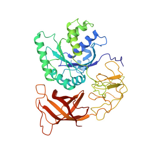
The crystal structure of elongation factor EF-Tu from Thermus aquaticus in the GTP conformation.

Kjeldgaard, M.Nissen, P.Thirup, S.Nyborg, J.

(1993) Structure 1: 35-50

  • DOI: https://doi.org/10.1016/0969-2126(93)90007-4
  • Primary Citation Related Structures: 
    1EFT

  • PubMed Abstract: 

    Elongation factor Tu (EF-Tu) is a GTP-binding protein that is crucial for protein biosynthesis. In the GTP form of the molecule, EF-Tu binds tightly to aminoacyl-tRNA, forming a ternary complex that interacts with the ribosomal acceptor site. During this interaction, GTP is hydrolyzed, and EF-Tu.GDP is ejected. The crystal structure of EF-Tu from Thermus aquaticus, complexed to the GTP analogue GDPNP, has been determined at 2.5 A resolution and compared to the structure of Escherichia coli EF-Tu.GDP. During the transition from the GDP (inactive) to the GTP (active) form, domain 1, containing the GTP-binding site, undergoes internal conformational changes similar to those observed in ras-p21. In addition, a dramatic rearrangement of domains is observed, corresponding to a rotation of 90.8 degrees of domain 1 relative to domains 2 and 3. Residues that are affected in the binding of aminoacyl-tRNA are found in or near the cleft formed by the domain interface. GTP binding by EF-Tu leads to dramatic conformational changes which expose the tRNA binding site. It appears that tRNA binding to EF-Tu induces a further conformational change, which may affect the GTPase activity.


  • Organizational Affiliation
    • Department of Chemistry, Aarhus University, Denmark.

Macromolecule Content 

  • Total Structure Weight: 45.28 kDa 
  • Atom Count: 3,302 
  • Modeled Residue Count: 405 
  • Deposited Residue Count: 405 
  • Unique protein chains: 1

Macromolecules

Find similar proteins by:|  3D Structure
Entity ID: 1
MoleculeChains  Sequence LengthOrganismDetailsImage
ELONGATION FACTOR TU405Thermus aquaticusMutation(s): 0 
EC: 3.6.5.3
UniProt
Find proteins for Q01698 (Thermus aquaticus)
Explore Q01698 
Go to UniProtKB:  Q01698
Entity Groups
Sequence Clusters30% Identity50% Identity70% Identity90% Identity95% Identity100% Identity
UniProt GroupQ01698
Sequence Annotations
Expand
Reference Sequence

Small Molecules

Ligands 2 Unique
IDChains Name / Formula / InChI Key2D Diagram3D Interactions
GNP

Query on GNP


Download:Ideal Coordinates CCD File
C [auth A]PHOSPHOAMINOPHOSPHONIC ACID-GUANYLATE ESTER
C10 H17 N6 O13 P3
UQABYHGXWYXDTK-UUOKFMHZSA-N
MG

Query on MG


Download:Ideal Coordinates CCD File
B [auth A]MAGNESIUM ION
Mg
JLVVSXFLKOJNIY-UHFFFAOYSA-N

Experimental Data & Validation

Experimental Data

  • Method: X-RAY DIFFRACTION
  • Resolution: 2.50 Å
  • R-Value Work:  0.200 (Depositor), 0.187 (DCC) 
  • R-Value Observed: 0.200 (Depositor) 
Space Group: C 1 2 1
Unit Cell:
Length ( Å )Angle ( ˚ )
a = 150.7α = 90
b = 99.7β = 95.3
c = 40.2γ = 90
Software Package:
Software NamePurpose
X-PLORmodel building
X-PLORrefinement
X-PLORphasing

Structure Validation

View Full Validation Report


Ligand Structure Quality Assessment 


Entry History 

Deposition Data

Revision History  (Full details and data files)

  • Version 1.0: 1994-08-31
    Type: Initial release
  • Version 1.1: 2008-03-24
    Changes: Version format compliance
  • Version 1.2: 2011-07-13
    Changes: Version format compliance
  • Version 1.3: 2024-02-07
    Changes: Data collection, Database references, Derived calculations, Other