1EFO | pdb_00001efo

CRYSTAL STRUCTURE OF AN ADENINE BULGE IN THE RNA CHAIN OF A DNA/RNA HYBRID, D(CTCCTCTTC)/R(GAAGAGAGAG)


Experimental Data Snapshot

  • Method: X-RAY DIFFRACTION
  • Resolution: 2.30 Å
  • R-Value Free: 
    0.222 (Depositor), 0.228 (DCC) 
  • R-Value Work: 
    0.199 (Depositor), 0.201 (DCC) 
  • R-Value Observed: 
    0.199 (Depositor) 

wwPDB Validation 3D Report Full Report

Validation slider image for 1EFO

This is version 1.4 of the entry. See complete history

Literature

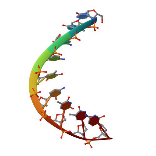
Crystal structure of an adenine bulge in the RNA chain of a DNA.RNA hybrid, d(CTCCTCTTC).r(gaagagagag).

Sudarsanakumar, C.Xiong, Y.Sundaralingam, M.

(2000) J Mol Biology 299: 103-112

  • DOI: https://doi.org/10.1006/jmbi.2000.3730
  • Primary Citation Related Structures: 
    1EFO

  • PubMed Abstract: 

    Crystal structure of a DNA.RNA hybrid, d(CTCCTCTTC).r(gaagagagag), with an adenine bulge in the polypurine RNA strand was determined at 2.3 A resolution. The structure was solved by the molecular replacement method and refined to a final R-factor of 19.9% (Rfree 22.2%). The hybrid duplex crystallized in the space group I222 with unit cell dimensions, a = 46.66 A, b = 47.61 A and c = 54.05 A, and adopts the A-form conformation. All RNA and DNA sugars are in the C3'-endo conformation, the glycosyl angles in anti conformation and the majority of the C4'-C5' torsion angles in g+ except two trans angles, in conformity with the C3'-endo rigid nucleotide hypothesis. The adenine bulge is looped out and it is also in the anti C3'-endo conformation. The bulge is involved in a base-triple (C.g)*a interaction with the end base-pair (C9.g10) in the minor groove of a symmetry-related molecule. The 2' hydroxyl group of g15 is hydrogen bonded to O2P and O5' of g17, skipping the bulged adenine a16 and stabilizing the sugar-phosphate backbone of the hybrid. The hydrogen bonding and the backbone conformation at the bulged adenine site is very similar to that found in the crystal structure of a protein-RNA complex.


  • Organizational Affiliation
    • Department of Chemistry, Biochemistry, Ohio State University, Columbus 43210, USA.

Macromolecule Content 

  • Total Structure Weight: 5.94 kDa 
  • Atom Count: 417 
  • Modeled Residue Count: 19 
  • Deposited Residue Count: 19 
  • Unique nucleic acid chains: 2

Macromolecules

Find similar nucleic acids by:  Sequence
Entity ID: 1
MoleculeChains LengthOrganismImage
DNA (5'-D(*CP*TP*CP*CP*TP*CP*TP*TP*C)-3')9N/A
Sequence Annotations
Expand
Reference Sequence
Find similar nucleic acids by:  Sequence
Entity ID: 2
MoleculeChains LengthOrganismImage
RNA (5'-R(*GP*AP*AP*GP*AP*GP*AP*GP*AP*G)-3')10N/A
Sequence Annotations
Expand
Reference Sequence

Experimental Data & Validation

Experimental Data

  • Method: X-RAY DIFFRACTION
  • Resolution: 2.30 Å
  • R-Value Free:  0.222 (Depositor), 0.228 (DCC) 
  • R-Value Work:  0.199 (Depositor), 0.201 (DCC) 
  • R-Value Observed: 0.199 (Depositor) 
Space Group: I 2 2 2
Unit Cell:
Length ( Å )Angle ( ˚ )
a = 46.66α = 90
b = 47.61β = 90
c = 54.04γ = 90
Software Package:
Software NamePurpose
AMoREphasing
CNSrefinement
SCALEPACKdata scaling

Structure Validation

View Full Validation Report


Entry History 

Deposition Data

Revision History  (Full details and data files)

  • Version 1.0: 2000-05-29
    Type: Initial release
  • Version 1.1: 2008-04-27
    Changes: Version format compliance
  • Version 1.2: 2011-07-13
    Changes: Version format compliance
  • Version 1.3: 2017-10-04
    Changes: Refinement description
  • Version 1.4: 2024-02-07
    Changes: Data collection, Database references