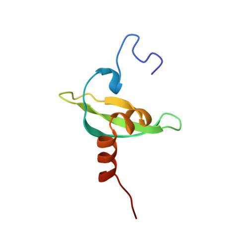
The Structure of Mouse Hp1 Suggests a Unique Mode of Single Peptide Recognition by the Shadow Chromo Domain Dimer
Brasher, S.V., Smith, B.O., Fogh, R.H., Nietlispach, D., Thiru, A., Nielsen, P.R., Broadhurst, R.W., Ball, L.J., Murzina, N., Laue, E.D.(2000) EMBO J 19: 1587
- PubMed: 10747027 Search on PubMedSearch on PubMed Central
- DOI: https://doi.org/10.1093/emboj/19.7.1587
- Primary Citation Related Structures:
1DZ1 - PubMed Abstract:
The heterochromatin protein 1 (HP1) family of proteins is involved in gene silencing via the formation of heterochromatic structures. They are composed of two related domains: an N-terminal chromo domain and a C-terminal shadow chromo domain. Present results suggest that chromo domains may function as protein interaction motifs, bringing together different proteins in multi-protein complexes and locating them in heterochromatin. We have previously determined the structure of the chromo domain from the mouse HP1beta protein, MOD1. We show here that, in contrast to the chromo domain, the shadow chromo domain is a homodimer. The intact HP1beta protein is also dimeric, where the interaction is mediated by the shadow chromo domain, with the chromo domains moving independently of each other at the end of flexible linkers. Mapping studies, with fragments of the CAF1 and TIF1beta proteins, show that an intact, dimeric, shadow chromo domain structure is required for complex formation.
- Cambridge Centre for Molecular Recognition, Department of Biochemistry, University of Cambridge, 80 Tennis Court Road, Cambridge CB2 1GA, UK.
Organizational Affiliation: