1DX5 | pdb_00001dx5

Crystal structure of the thrombin-thrombomodulin complex


Experimental Data Snapshot

  • Method: X-RAY DIFFRACTION
  • Resolution: 2.30 Å
  • R-Value Free: 
    0.241 (Depositor) 
  • R-Value Work: 
    0.200 (Depositor) 
  • R-Value Observed: 
    0.200 (Depositor) 

Starting Model: other
View more details

wwPDB Validation 3D Report Full Report

Validation slider image for 1DX5

This is version 2.3 of the entry. See complete history

Literature

Structural Basis for the Anticoagulant Activity of the Thrombin-Thrombomodulin Complex

Fuentes-Prior, P.Iwanaga, Y.Huber, R.Pagila, R.Rumennik, G.Seto, M.Morser, J.Light, D.R.Bode, W.

(2000) Nature 404: 518

  • DOI: https://doi.org/10.1038/35006683
  • Primary Citation Related Structures: 
    1DX5

  • PubMed Abstract: 

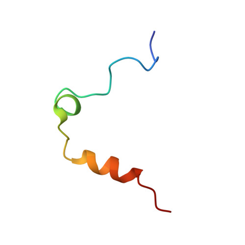
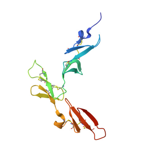
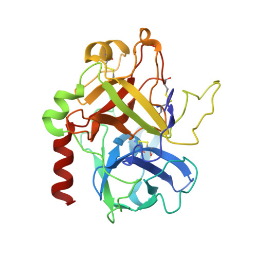
    The serine proteinase alpha-thrombin causes blood clotting through proteolytic cleavage of fibrinogen and protease-activated receptors and amplifies its own generation by activating the essential clotting factors V and VIII. Thrombomodulin, a transmembrane thrombin receptor with six contiguous epidermal growth factor-like domains (TME1-6), profoundly alters the substrate specificity of thrombin from pro- to anticoagulant by activating protein C. Activated protein C then deactivates the coagulation cascade by degrading activated factors V and VIII. The thrombin-thrombomodulin complex inhibits fibrinolysis by activating the procarboxypeptidase thrombin-activatable fibrinolysis inhibitor. Here we present the 2.3 A crystal structure of human alpha-thrombin bound to the smallest thrombomodulin fragment required for full protein-C co-factor activity, TME456. The Y-shaped thrombomodulin fragment binds to thrombin's anion-binding exosite-I, preventing binding of procoagulant substrates. Thrombomodulin binding does not seem to induce marked allosteric structural rearrangements at the thrombin active site. Rather, docking of a protein C model to thrombin-TME456 indicates that TME45 may bind substrates in such a manner that their zymogen-activation cleavage sites are presented optimally to the unaltered thrombin active site.


  • Organizational Affiliation
    • Max-Planck-Institut für Biochemie, Martinsried, Germany. fuentes@biochem.mpg.de

Macromolecule Content 

  • Total Structure Weight: 194.11 kDa 
  • Atom Count: 14,018 
  • Modeled Residue Count: 1,652 
  • Deposited Residue Count: 1,652 
  • Unique protein chains: 3

Macromolecules

Find similar proteins by:|  3D Structure
Entity ID: 1
MoleculeChains  Sequence LengthOrganismDetailsImage
Thrombin light chain
A, B, C, D
36Homo sapiensMutation(s): 0 
EC: 3.4.21.5
UniProt & NIH Common Fund Data Resources
Find proteins for P00734 (Homo sapiens)
Explore P00734 
Go to UniProtKB:  P00734
PHAROS:  P00734
GTEx:  ENSG00000180210 
Entity Groups
Sequence Clusters30% Identity50% Identity70% Identity90% Identity95% Identity100% Identity
UniProt GroupP00734
Sequence Annotations
Expand
Reference Sequence
Find similar proteins by:|  3D Structure
Entity ID: 2
MoleculeChains  Sequence LengthOrganismDetailsImage
ThrombomodulinE [auth I],
F [auth J],
G [auth K],
H [auth L]
118Homo sapiensMutation(s): 0 
Gene Names: THBDTHRM
UniProt & NIH Common Fund Data Resources
Find proteins for P07204 (Homo sapiens)
Explore P07204 
Go to UniProtKB:  P07204
PHAROS:  P07204
GTEx:  ENSG00000178726 
Entity Groups
Sequence Clusters30% Identity50% Identity70% Identity90% Identity95% Identity100% Identity
UniProt GroupP07204
Sequence Annotations
Expand
Reference Sequence
Find similar proteins by:|  3D Structure
Entity ID: 3
MoleculeChains  Sequence LengthOrganismDetailsImage
Thrombin heavy chainI [auth M],
J [auth N],
K [auth O],
L [auth P]
259Homo sapiensMutation(s): 0 
EC: 3.4.21.5
UniProt & NIH Common Fund Data Resources
Find proteins for P00734 (Homo sapiens)
Explore P00734 
Go to UniProtKB:  P00734
PHAROS:  P00734
GTEx:  ENSG00000180210 
Entity Groups
Sequence Clusters30% Identity50% Identity70% Identity90% Identity95% Identity100% Identity
UniProt GroupP00734
Glycosylation
Glycosylation Sites: 1Go to GlyGen: P00734-1
Sequence Annotations
Expand
Reference Sequence

Small Molecules

Ligands 5 Unique
IDChains Name / Formula / InChI Key2D Diagram3D Interactions
0GJ

Query on 0GJ


Download:Ideal Coordinates CCD File
AA [auth M]
BA [auth M]
CA [auth M]
DA [auth M]
GA [auth N]
AA [auth M],
BA [auth M],
CA [auth M],
DA [auth M],
GA [auth N],
HA [auth N],
IA [auth N],
JA [auth N],
MA [auth O],
NA [auth O],
OA [auth O],
PA [auth O],
TA [auth P],
UA [auth P],
VA [auth P],
WA [auth P]
L-alpha-glutamyl-N-{(1S)-4-{[amino(iminio)methyl]amino}-1-[(1S)-2-chloro-1-hydroxyethyl]butyl}glycinamide
C14 H28 Cl N6 O5
XELWNHKFCNMWQO-LPEHRKFASA-O
NAG

Query on NAG


Download:Ideal Coordinates CCD File
EA [auth N],
KA [auth O],
QA [auth P],
Y [auth M]
2-acetamido-2-deoxy-beta-D-glucopyranose
C8 H15 N O6
OVRNDRQMDRJTHS-FMDGEEDCSA-N
FMT

Query on FMT


Download:Ideal Coordinates CCD File
M [auth I],
P [auth J],
S [auth K],
SA [auth P],
V [auth L]
FORMIC ACID
C H2 O2
BDAGIHXWWSANSR-UHFFFAOYSA-N
CA

Query on CA


Download:Ideal Coordinates CCD File
N [auth I],
Q [auth J],
T [auth K],
W [auth L]
CALCIUM ION
Ca
BHPQYMZQTOCNFJ-UHFFFAOYSA-N
NA

Query on NA


Download:Ideal Coordinates CCD File
FA [auth N]
LA [auth O]
O [auth I]
R [auth J]
RA [auth P]
FA [auth N],
LA [auth O],
O [auth I],
R [auth J],
RA [auth P],
U [auth K],
X [auth L],
Z [auth M]
SODIUM ION
Na
FKNQFGJONOIPTF-UHFFFAOYSA-N

Biologically Interesting Molecules (External Reference) 

1 Unique
Entity ID: 8
IDChains NameType/Class2D Diagram3D Interactions
PRD_000288 (0GJ)
Query on PRD_000288
AA [auth M]
BA [auth M]
CA [auth M]
DA [auth M]
GA [auth N]
AA [auth M], BA [auth M], CA [auth M], DA [auth M], GA [auth N], HA [auth N], IA [auth N], JA [auth N], MA [auth O], NA [auth O], OA [auth O], PA [auth O], TA [auth P], UA [auth P], VA [auth P], WA [auth P]
GLU-GLY-ARG-CHLOROMETHYL KETONEPeptide-like / Inhibitor

Experimental Data & Validation

Experimental Data

  • Method: X-RAY DIFFRACTION
  • Resolution: 2.30 Å
  • R-Value Free:  0.241 (Depositor) 
  • R-Value Work:  0.200 (Depositor) 
  • R-Value Observed: 0.200 (Depositor) 
Space Group: H 3
Unit Cell:
Length ( Å )Angle ( ˚ )
a = 214.4α = 90
b = 214.4β = 90
c = 131.41γ = 120
Software Package:
Software NamePurpose
X-PLORrefinement
MOSFLMdata reduction
SCALAdata scaling
X-PLORphasing

Structure Validation

View Full Validation Report


Entry History 

Deposition Data

Revision History  (Full details and data files)

  • Version 1.0: 2000-04-10
    Type: Initial release
  • Version 1.1: 2011-07-13
    Changes: Atomic model, Database references, Derived calculations, Non-polymer description, Structure summary, Version format compliance
  • Version 1.2: 2016-12-21
    Changes: Advisory, Atomic model, Derived calculations, Non-polymer description, Other, Source and taxonomy
  • Version 2.0: 2018-06-27
    Changes: Advisory, Atomic model, Data collection, Database references, Derived calculations, Non-polymer description, Polymer sequence, Source and taxonomy, Structure summary
  • Version 2.1: 2020-07-29
    Type: Remediation
    Reason: Carbohydrate remediation
    Changes: Data collection, Derived calculations, Structure summary
  • Version 2.2: 2024-05-01
    Changes: Advisory, Data collection, Database references, Refinement description, Structure summary
  • Version 2.3: 2024-11-13
    Changes: Structure summary