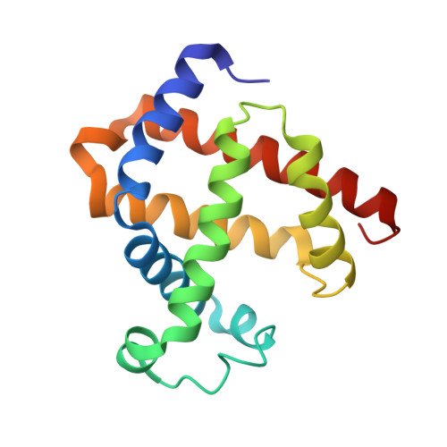
1DUK

WILD-TYPE RECOMBINANT SPERM WHALE METAQUOMYOGLOBIN


Experimental Data Snapshot

  • Method: X-RAY DIFFRACTION
  • Resolution: 2.13 Å
  • R-Value Observed: 0.180 

wwPDB Validation   3D Report Full Report


Ligand Structure Quality Assessment 


This is version 1.3 of the entry. See complete history


Literature

Trans-substitution of the proximal hydrogen bond in myoglobin: I. Structural consequences of hydrogen bond deletion.

Barrick, D.Dahlquist, F.W.

(2000) Proteins 39: 278-290

  • DOI: https://doi.org/10.1002/(sici)1097-0134(20000601)39:4<278::aid-prot20>3.0.co;2-t
  • Primary Citation of Related Structures:  
    1DTM, 1DUK, 1DUO

  • PubMed Abstract: 

    The structural role of a side-chain to side-chain protein hydrogen bond is examined using trans-substitution of the proximal histidine of myoglobin with methylimidazoles (Barrick, Biochemistry 1994;33:6546-6554). Modification of the chemical structure of exogenous ligands allows this hydrogen bond to be disrupted. Comparison of the crystal structures of H93G myoglobin complexed 4-methylimidazole (4meimd; methylation at carbon 4) and 1-methylimidazole (1meimd; methylation at the adjacent nitrogen, preventing hydrogen bonding between the imidazole ligand and the protein) shows that the polypeptide, heme, and methylimidazole orientations are the same within error. For 4meimd there appear to be major and minor conformations corresponding to different tautomeric states of the ligand. Conformational heterogeneity is also seen in the hyperfine-shifted region of the NMR spectrum of 4meimd complexed with high-spin H93G deoxyMb. The major conformation of the 4meimd ligand and the 1meimd ligand, as seen in the respective crystal structures, are quite similar except that the proximal ligand NH-to-Ser92-OH hydrogen bond is eliminated in the 1meimd complex, and instead the proximal ligand CH is adjacent to the Ser92-OH. Thus, this system provides a means to eliminate the Mb proximal hydrogen bond in a chemically and structurally conservative way.


  • Organizational Affiliation

    The T.C. Jenkins Department of Biophysics, Johns Hopkins University, Baltimore, Maryland 21218, USA. barrick@jhu.edu


Macromolecules
Find similar proteins by:  (by identity cutoff)  |  3D Structure
Entity ID: 1
MoleculeChains Sequence LengthOrganismDetailsImage
WILD-TYPE RECOMBINANT SPERM WHALE METAQUOMYOGLOBIN153Physeter catodonMutation(s): 0 
EC: 1.11.1 (UniProt), 1.7 (UniProt)
UniProt
Find proteins for P02185 (Physeter macrocephalus)
Explore P02185 
Go to UniProtKB:  P02185
Entity Groups  
Sequence Clusters30% Identity50% Identity70% Identity90% Identity95% Identity100% Identity
UniProt GroupP02185
Sequence Annotations
Expand
  • Reference Sequence
Small Molecules
Ligands 1 Unique
IDChains Name / Formula / InChI Key2D Diagram3D Interactions
HEM
Query on HEM

Download Ideal Coordinates CCD File 
B [auth A]PROTOPORPHYRIN IX CONTAINING FE
C34 H32 Fe N4 O4
KABFMIBPWCXCRK-RGGAHWMASA-L
Experimental Data & Validation

Experimental Data

  • Method: X-RAY DIFFRACTION
  • Resolution: 2.13 Å
  • R-Value Observed: 0.180 
  • Space Group: P 21 21 21
Unit Cell:
Length ( Å )Angle ( ˚ )
a = 40.132α = 90
b = 49.071β = 90
c = 79.339γ = 90
Software Package:
Software NamePurpose
SDMSdata collection
SDMSdata reduction
TNTrefinement
SDMSdata scaling
TNTphasing

Structure Validation

View Full Validation Report


Ligand Structure Quality Assessment 


Entry History 

Deposition Data

Revision History  (Full details and data files)

  • Version 1.0: 2000-02-07
    Type: Initial release
  • Version 1.1: 2008-04-27
    Changes: Version format compliance
  • Version 1.2: 2011-07-13
    Changes: Version format compliance
  • Version 1.3: 2024-02-07
    Changes: Data collection, Database references, Derived calculations