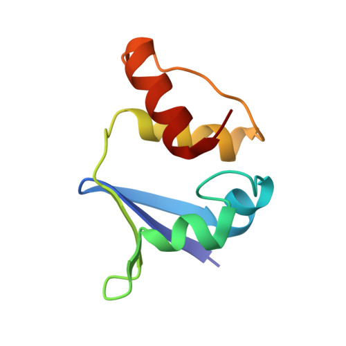
1DSX

KV1.2 T1 DOMAIN, RESIDUES 33-119, T46V MUTANT


Experimental Data Snapshot

  • Method: X-RAY DIFFRACTION
  • Resolution: 1.60 Å
  • R-Value Free: 0.279 
  • R-Value Work: 0.237 

wwPDB Validation   3D Report Full Report


This is version 1.4 of the entry. See complete history


Literature

The polar T1 interface is linked to conformational changes that open the voltage-gated potassium channel.

Minor, D.L.Lin, Y.F.Mobley, B.C.Avelar, A.Jan, Y.N.Jan, L.Y.Berger, J.M.

(2000) Cell 102: 657-670

  • DOI: https://doi.org/10.1016/s0092-8674(00)00088-x
  • Primary Citation of Related Structures:  
    1DSX, 1QDV, 1QDW

  • PubMed Abstract: 

    Kv voltage-gated potassium channels share a cytoplasmic assembly domain, T1. Recent mutagenesis of two T1 C-terminal loop residues implicates T1 in channel gating. However, structural alterations of these mutants leave open the question concerning direct involvement of T1 in gating. We find in mammalian Kv1.2 that gating depends critically on residues at complementary T1 surfaces in an unusually polar interface. An isosteric mutation in this interface causes surprisingly little structural alteration while stabilizing the closed channel and increasing the stability of T1 tetramers. Replacing T1 with a tetrameric coiled-coil destabilizes the closed channel. Together, these data suggest that structural changes involving the buried polar T1 surfaces play a key role in the conformational changes leading to channel opening.


  • Organizational Affiliation

    Howard Hughes Medical Institute and Department of Physiology, University of California, San Francisco 94143, USA. minor@itsa.ucsf.edu


Macromolecules
Find similar proteins by:  (by identity cutoff)  |  3D Structure
Entity ID: 1
MoleculeChains Sequence LengthOrganismDetailsImage
PROTEIN (KV1.2 VOLTAGE-GATED POTASSIUM CHANNEL)
A, B, C, D, E
A, B, C, D, E, F, G, H
87Rattus norvegicusMutation(s): 1 
UniProt
Find proteins for P63142 (Rattus norvegicus)
Explore P63142 
Go to UniProtKB:  P63142
Entity Groups  
Sequence Clusters30% Identity50% Identity70% Identity90% Identity95% Identity100% Identity
UniProt GroupP63142
Sequence Annotations
Expand
  • Reference Sequence
Experimental Data & Validation

Experimental Data

  • Method: X-RAY DIFFRACTION
  • Resolution: 1.60 Å
  • R-Value Free: 0.279 
  • R-Value Work: 0.237 
  • Space Group: P 21 21 21
Unit Cell:
Length ( Å )Angle ( ˚ )
a = 76.39α = 90
b = 78.398β = 90
c = 126.924γ = 90
Software Package:
Software NamePurpose
AMoREphasing
REFMACrefinement
DENZOdata reduction
SCALEPACKdata scaling

Structure Validation

View Full Validation Report


Entry History 

Deposition Data

Revision History  (Full details and data files)

  • Version 1.0: 2000-09-20
    Type: Initial release
  • Version 1.1: 2008-04-27
    Changes: Version format compliance
  • Version 1.2: 2011-07-13
    Changes: Version format compliance
  • Version 1.3: 2021-11-03
    Changes: Database references
  • Version 1.4: 2024-02-07
    Changes: Data collection