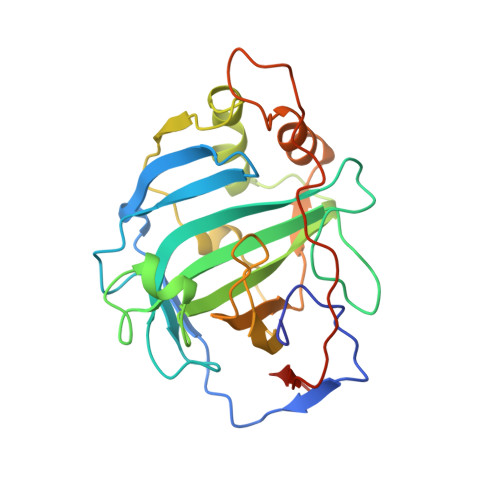
Structure determination of murine mitochondrial carbonic anhydrase V at 2.45-A resolution: implications for catalytic proton transfer and inhibitor design.
Boriack-Sjodin, P.A., Heck, R.W., Laipis, P.J., Silverman, D.N., Christianson, D.W.(1995) Proc Natl Acad Sci U S A 92: 10949-10953
- PubMed: 7479916
- DOI: https://doi.org/10.1073/pnas.92.24.10949
- Primary Citation of Related Structures:
1DMX, 1DMY - PubMed Abstract:
The three-dimensional structure of murine mitochondrial carbonic anhydrase V has been determined and refined at 2.45-A resolution (crystallographic R factor = 0.187). Significant structural differences unique to the active site of carbonic anhydrase V are responsible for differences in the mechanism of catalytic proton transfer as compared with other carbonic anhydrase isozymes. In the prototypical isozyme, carbonic anhydrase II, catalytic proton transfer occurs via the shuttle group His-64; carbonic anhydrase V has Tyr-64, which is not an efficient proton shuttle due in part to the bulky adjacent side chain of Phe-65. Based on analysis of the structure of carbonic anhydrase V, we speculate that Tyr-131 may participate in proton transfer due to its proximity to zinc-bound solvent, its solvent accessibility, and its electrostatic environment in the protein structure. Finally, the design of isozyme-specific inhibitors is discussed in view of the complex between carbonic anhydrase V and acetazolamide, a transition-state analogue. Such inhibitors may be physiologically important in the regulation of blood glucose levels.
- Department of Chemistry, University of Pennsylvania, Philadelphia 19104-6323, USA.
Organizational Affiliation:


