Acridinecarboxamide topoisomerase poisons: structural and kinetic studies of the DNA complexes of 5-substituted 9-amino-(N-(2-dimethylamino)ethyl)acridine-4-carboxamides.
Adams, A., Guss, J.M., Collyer, C.A., Denny, W.A., Prakash, A.S., Wakelin, L.P.(2000) Mol Pharmacol 58: 649-658
- PubMed: 10953060 Search on PubMed
- DOI: https://doi.org/10.1124/mol.58.3.649
- Primary Citation Related Structures:
1DL8 - PubMed Abstract:
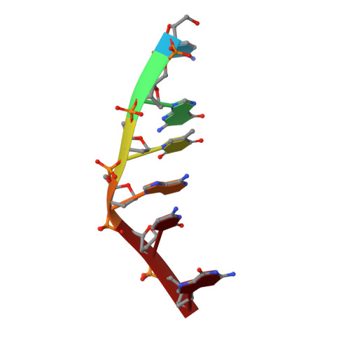
For a series of antitumor-active 5-substituted 9-aminoacridine-4-carboxamide topoisomerase II poisons, we have used X-ray crystallography and stopped-flow spectrophotometry to explore relationships between DNA binding kinetics, biological activity, and the structures of their DNA complexes. The structure of 5-F-9-amino-[N-(2-dimethylamino)ethyl]-acridine-4-carboxamide bound to d(CGTACG)(2) has been solved to a resolution of 1.55 A in space group P6(4). A drug molecule intercalates between each of the CpG dinucleotide steps, its protonated dimethylamino group partially occupying positions close to the N7 and O6 atoms of guanine G2 in the major groove. A water molecule forms bridging hydrogen bonds between the 4-carboxamide NH and the phosphate group of the same guanine. Intercalation unwinds steps 1 and 2 by 12 degrees and 8 degrees, respectively compared with B-DNA, whereas the central TpA step is overwound by 10 degrees. Nonphenyl 5-substituents, on average, decrease mean DNA dissociation rates by a factor of three, regardless of their steric, hydrophobic, H-bonding, or electronic properties. Cytotoxicity is enhanced on average 4-fold and binding affinities rise by 3-fold, thus there is an apparent association between kinetics, affinity, and cytotoxicity. Taken together, the structural and kinetic studies imply that the main origin of this association is enhanced stacking interactions between the 5-substituent and cytosine in the CpG binding site. Ligand-dependent perturbations in base pair twist angles and their consequent effects on base pair-base pair stacking interactions may also contribute to the stability of the intercalated complex. 5-Phenyl substituents modify dissociation rates without affecting affinities, and variations in their biological activity are not correlated with DNA binding properties, which suggests that they interact directly with the topoisomerase protein.
- Department of Biochemistry, University of Sydney, New South Wales, Australia. a.adams@biochem.usyd.edu.au
Organizational Affiliation: