1DK5 | pdb_00001dk5

CRYSTAL STRUCTURE OF ANNEXIN 24(CA32) FROM CAPSICUM ANNUUM


Experimental Data Snapshot

  • Method: X-RAY DIFFRACTION
  • Resolution: 2.80 Å
  • R-Value Free: 
    0.316 (Depositor) 
  • R-Value Work: 
    0.216 (Depositor), 0.228 (DCC) 
  • R-Value Observed: 
    0.216 (Depositor) 

wwPDB Validation 3D Report Full Report

Validation slider image for 1DK5

This is version 1.5 of the entry. See complete history

Literature

Annexin 24 from Capsicum annuum. X-ray structure and biochemical characterization.

Hofmann, A.Proust, J.Dorowski, A.Schantz, R.Huber, R.

(2000) J Biological Chem 275: 8072-8082

  • DOI: https://doi.org/10.1074/jbc.275.11.8072
  • Primary Citation Related Structures: 
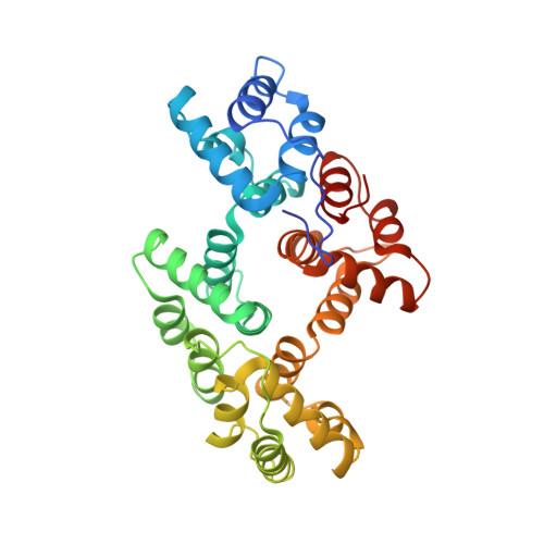
    1DK5

  • PubMed Abstract: 

    This work provides the first three-dimensional structure of a member of the plant annexin family and correlates these findings with biochemical properties of this protein. Annexin 24(Ca32) from Capsicum annuum was purified as a native protein from bell pepper and was also prepared by recombinant techniques. To overcome the problem of precipitation of the recombinant wild-type protein in crystallization trials, two mutants were designed. Whereas an N-terminal truncation mutant turned out to be an unstable protein, the N-terminal His-tagged annexin 24(Ca32) was crystallized, and the three-dimensional structure was determined by x-ray diffraction at 2. 8 A resolution. The structure refined to an R-factor of 0.216 adopts the typical annexin fold; the detailed structure, however, is different from non-plant annexins, especially in domains I and III and in the membrane binding loops on the convex side. Within the unit cell there are two molecules per asymmetric unit, which differ in conformation of the IAB-loop. Both conformers show Trp-35 on the surface. The loop-out conformation is stabilized by tight interactions of this tryptophan with residue side chains of a symmetry-related molecule and enforced by a bound sulfate. Characterization of this plant annexin using biophysical methods revealed calcium-dependent binding to phospholipid vesicles with preference for phosphatidylcholine over phosphatidylserine and magnesium-dependent phosphodiesterase activity in vitro as shown with adenosine triphosphate as the substrate. A comparative unfolding study of recombinant annexin 24(Ca32) wild type and of the His-tag fusion protein indicates higher stability of the latter. The effect of this N-terminal modification is also visible from CD spectra. Both proteins were subjected to a FURA-2-based calcium influx assay, which gave high influx rates for the wild-type but greatly reduced influx rates for the fusion protein. We therefore conclude that the N-terminal domain is indeed a major regulatory element modulating different annexin properties by allosteric mechanisms.


  • Organizational Affiliation
    • Max-Planck-Institut für Biochemie, Abt. Strukturforschung, 82152 Martinsried, Germany. hofmanna@ncifcrf.gov

Macromolecule Content 

  • Total Structure Weight: 77.05 kDa 
  • Atom Count: 5,572 
  • Modeled Residue Count: 630 
  • Deposited Residue Count: 644 
  • Unique protein chains: 1

Macromolecules

Find similar proteins by:|  3D Structure
Entity ID: 1
MoleculeChains  Sequence LengthOrganismDetailsImage
ANNEXIN 24(CA32)
A, B
322Capsicum annuumMutation(s): 0 
UniProt
Find proteins for Q42657 (Capsicum annuum)
Explore Q42657 
Go to UniProtKB:  Q42657
Entity Groups
Sequence Clusters30% Identity50% Identity70% Identity90% Identity95% Identity100% Identity
UniProt GroupQ42657
Sequence Annotations
Expand
Reference Sequence

Small Molecules

Ligands 1 Unique
IDChains Name / Formula / InChI Key2D Diagram3D Interactions
SO4

Query on SO4


Download:Ideal Coordinates CCD File
AA [auth B]
BA [auth B]
C [auth A]
CA [auth B]
D [auth A]
AA [auth B],
BA [auth B],
C [auth A],
CA [auth B],
D [auth A],
DA [auth B],
E [auth A],
EA [auth B],
F [auth A],
FA [auth B],
G [auth A],
GA [auth B],
H [auth A],
HA [auth B],
I [auth A],
IA [auth B],
J [auth A],
K [auth A],
L [auth A],
M [auth A],
N [auth A],
O [auth A],
P [auth A],
Q [auth B],
R [auth B],
S [auth B],
T [auth B],
U [auth B],
V [auth B],
W [auth B],
X [auth B],
Y [auth B],
Z [auth B]
SULFATE ION
O4 S
QAOWNCQODCNURD-UHFFFAOYSA-L

Experimental Data & Validation

Experimental Data

  • Method: X-RAY DIFFRACTION
  • Resolution: 2.80 Å
  • R-Value Free:  0.316 (Depositor) 
  • R-Value Work:  0.216 (Depositor), 0.228 (DCC) 
  • R-Value Observed: 0.216 (Depositor) 
Space Group: P 32 2 1
Unit Cell:
Length ( Å )Angle ( ˚ )
a = 96.76α = 90
b = 96.76β = 90
c = 173.39γ = 120
Software Package:
Software NamePurpose
AMoREphasing
CNSrefinement
MOSFLMdata reduction
CCP4data scaling

Structure Validation

View Full Validation Report


Entry History 

Deposition Data

Revision History  (Full details and data files)

  • Version 1.0: 2000-12-06
    Type: Initial release
  • Version 1.1: 2008-04-27
    Changes: Version format compliance
  • Version 1.2: 2011-07-13
    Changes: Derived calculations, Version format compliance
  • Version 1.3: 2017-12-20
    Changes: Database references
  • Version 1.4: 2018-01-31
    Changes: Experimental preparation
  • Version 1.5: 2024-02-07
    Changes: Data collection, Database references, Derived calculations