1DG6

CRYSTAL STRUCTURE OF APO2L/TRAIL


Experimental Data Snapshot

  • Method: X-RAY DIFFRACTION
  • Resolution: 1.30 Å
  • R-Value Free: 0.197 
  • R-Value Work: 0.141 
  • R-Value Observed: 0.141 

wwPDB Validation   3D Report Full Report


This is version 1.4 of the entry. See complete history


Literature

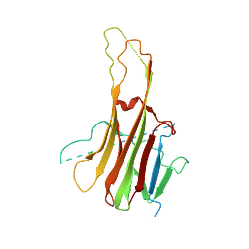
A unique zinc-binding site revealed by a high-resolution X-ray structure of homotrimeric Apo2L/TRAIL.

Hymowitz, S.G.O'Connell, M.P.Ultsch, M.H.Hurst, A.Totpal, K.Ashkenazi, A.de Vos, A.M.Kelley, R.F.

(2000) Biochemistry 39: 633-650

  • DOI: https://doi.org/10.1021/bi992242l
  • Primary Citation of Related Structures:  
    1DG6

  • PubMed Abstract: 

    Apoptosis-inducing ligand 2 (Apo2L, also called TRAIL), a member of the tumor necrosis factor (TNF) family, induces apoptosis in a variety of human tumor cell lines but not in normal cells [Wiley, S. R., Schooley, K., Smolak, P. J., Din, W. S., Huang, C.-P., Nicholl, J. K., Sutherland, G. R., Smith, T. D., Rauch, C., Smith, C. A., and Goodwin, R. G. (1995) Immunity 3, 673-682; Pitti, R. M., Marsters, S. A., Ruppert, S., Donahue, C. J., Moore, A., and Ashkenazi, A. (1996) J. Biol. Chem. 271, 12687-12690]. Here we describe the structure of Apo2L at 1.3 A resolution and use alanine-scanning mutagenesis to map the receptor contact regions. The structure reveals a homotrimeric protein that resembles TNF with receptor-binding epitopes at the interface between monomers. A zinc ion is buried at the trimer interface, coordinated by the single cysteine residue of each monomer. The zinc ion is required for maintaining the native structure and stability and, hence, the biological activity of Apo2L. This is the first example of metal-dependent oligomerization and function of a cytokine.


  • Organizational Affiliation

    Department of Protein Engineering, Genentech, Inc., 1 DNA Way, South San Francisco, California 94080, USA.


Macromolecules
Find similar proteins by:  (by identity cutoff)  |  3D Structure
Entity ID: 1
MoleculeChains Sequence LengthOrganismDetailsImage
APO2L/TNF-RELATED APOPOTIS INDUCING LIGAND (TRAIL)191Homo sapiensMutation(s): 1 
UniProt & NIH Common Fund Data Resources
Find proteins for P50591 (Homo sapiens)
Explore P50591 
Go to UniProtKB:  P50591
PHAROS:  P50591
GTEx:  ENSG00000121858 
Entity Groups  
Sequence Clusters30% Identity50% Identity70% Identity90% Identity95% Identity100% Identity
UniProt GroupP50591
Sequence Annotations
Expand
  • Reference Sequence
Experimental Data & Validation

Experimental Data

  • Method: X-RAY DIFFRACTION
  • Resolution: 1.30 Å
  • R-Value Free: 0.197 
  • R-Value Work: 0.141 
  • R-Value Observed: 0.141 
  • Space Group: H 3 2
Unit Cell:
Length ( Å )Angle ( ˚ )
a = 66.435α = 90
b = 66.435β = 90
c = 197.727γ = 120
Software Package:
Software NamePurpose
AMoREphasing
SHELXL-97refinement
DENZOdata reduction
SCALEPACKdata scaling

Structure Validation

View Full Validation Report


Entry History 

Deposition Data

Revision History  (Full details and data files)

  • Version 1.0: 2000-01-26
    Type: Initial release
  • Version 1.1: 2008-04-27
    Changes: Version format compliance
  • Version 1.2: 2011-07-13
    Changes: Derived calculations, Version format compliance
  • Version 1.3: 2018-03-14
    Changes: Database references
  • Version 1.4: 2023-08-09
    Changes: Data collection, Database references, Derived calculations, Refinement description