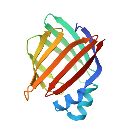
Properties and crystal structure of a beta-barrel folding mutant.
Ropson, I.J., Yowler, B.C., Dalessio, P.M., Banaszak, L., Thompson, J.(2000) Biophys J 78: 1551-1560
- PubMed: 10692339
- DOI: https://doi.org/10.1016/S0006-3495(00)76707-5
- Primary Citation of Related Structures:
1DC9 - PubMed Abstract:
A mutant of a beta-barrel protein, rat intestinal fatty acid binding protein, was predicted to be more stable than the wild-type protein due to a novel hydrogen bond. Equilibrium denaturation studies indicated the opposite: the V60N mutant protein was less stable. The folding transitions followed by CD and fluorescence were reversible and two-state for both mutant and wild-type protein. However, the rates of denaturation and renaturation of V60N were faster. During unfolding, the initial rate was associated with 75-80% of the fluorescence and all of the CD amplitude change. A subsequent rate accounted for the remaining fluorescence change for both proteins; thus the intermediate state lacked secondary structure. During folding, one rate was detected by both fluorescence and CD after an initial burst phase for both wild-type and mutant. An additional slower folding rate was detected by fluorescence for the mutant protein. The structure of the V60N mutant has been obtained and is nearly identical to prior crystal structures of IFABP. Analysis of mean differences in hydrogen bond and van der Waals interactions did not readily account for the stability loss due to the mutation. However, significant average differences of the solvent accessible surface and crystallographic displacement factors suggest entropic destabilization.
- Department of Biochemistry and Molecular Biology, Penn State University College of Medicine, Hershey, Pennsylvania 17033, USA.
Organizational Affiliation:


