1DAM

DETHIOBIOTIN SYNTHETASE COMPLEXED WITH DETHIOBIOTIN, ADP, INORGANIC PHOSPHATE AND MAGNESIUM


Experimental Data Snapshot

  • Method: X-RAY DIFFRACTION
  • Resolution: 1.80 Å
  • R-Value Free: 0.233 
  • R-Value Work: 0.180 

wwPDB Validation   3D Report Full Report


Ligand Structure Quality Assessment 


This is version 1.5 of the entry. See complete history


Literature

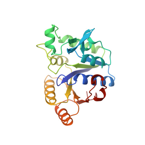
Crystal structure of two quaternary complexes of dethiobiotin synthetase, enzyme-MgADP-AlF3-diaminopelargonic acid and enzyme-MgADP-dethiobiotin-phosphate; implications for catalysis.

Kack, H.Sandmark, J.Gibson, K.J.Schneider, G.Lindqvist, Y.

(1998) Protein Sci 7: 2560-2566

  • DOI: https://doi.org/10.1002/pro.5560071209
  • Primary Citation of Related Structures:  
    1BS1, 1DAM

  • PubMed Abstract: 

    The crystal structures of two complexes of dethiobiotin synthetase, enzyme-diaminopelargonic acid-MgADP-AlF3 and enzyme-dethiobiotin-MgADP-Pi, respectively, have been determined to 1.8 A resolution. In dethiobiotin synthetase, AlF3 together with carbamylated diaminopelargonic acid mimics the phosphorylated reaction intermediate rather than the transition state complex for phosphoryl transfer. Observed differences in the binding of substrate, diaminopelargonic acid, and the product, dethiobiotin, suggest considerable displacements of substrate atoms during the ring closure step of the catalytic reaction. In both complexes, two metal ions are observed at the active site, providing evidence for a two-metal mechanism for this enzyme.


  • Organizational Affiliation

    Department of Medical Biochemistry and Biophysics, Karolinska Institutet, Stockholm, Sweden.


Macromolecules
Find similar proteins by:  (by identity cutoff)  |  3D Structure
Entity ID: 1
MoleculeChains Sequence LengthOrganismDetailsImage
PROTEIN (DETHIOBIOTIN SYNTHETASE)224Escherichia coliMutation(s): 0 
EC: 6.3.3.3
UniProt
Find proteins for P13000 (Escherichia coli (strain K12))
Explore P13000 
Go to UniProtKB:  P13000
Entity Groups  
Sequence Clusters30% Identity50% Identity70% Identity90% Identity95% Identity100% Identity
UniProt GroupP13000
Sequence Annotations
Expand
  • Reference Sequence
Experimental Data & Validation

Experimental Data

  • Method: X-RAY DIFFRACTION
  • Resolution: 1.80 Å
  • R-Value Free: 0.233 
  • R-Value Work: 0.180 
  • Space Group: C 1 2 1
Unit Cell:
Length ( Å )Angle ( ˚ )
a = 72.7α = 90
b = 48.9β = 106.7
c = 61.1γ = 90
Software Package:
Software NamePurpose
REFMACrefinement
DENZOdata reduction
SCALEPACKdata scaling

Structure Validation

View Full Validation Report


Ligand Structure Quality Assessment 


Entry History 

Deposition Data

Revision History  (Full details and data files)

  • Version 1.0: 1999-01-13
    Type: Initial release
  • Version 1.1: 2008-04-27
    Changes: Version format compliance
  • Version 1.2: 2011-07-13
    Changes: Derived calculations, Version format compliance
  • Version 1.3: 2016-04-06
    Changes: Non-polymer description
  • Version 1.4: 2017-10-04
    Changes: Refinement description
  • Version 1.5: 2024-02-07
    Changes: Data collection, Database references, Derived calculations