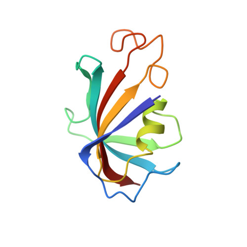
1D7H

FKBP COMPLEXED WITH DMSO


Experimental Data Snapshot

  • Method: X-RAY DIFFRACTION
  • Resolution: 1.90 Å
  • R-Value Free: 0.273 
  • R-Value Work: 0.213 
  • R-Value Observed: 0.213 

wwPDB Validation   3D Report Full Report


This is version 1.4 of the entry. See complete history


Literature

X-ray structures of small ligand-FKBP complexes provide an estimate for hydrophobic interaction energies.

Burkhard, P.Taylor, P.Walkinshaw, M.D.

(2000) J Mol Biol 295: 953-962

  • DOI: https://doi.org/10.1006/jmbi.1999.3411
  • Primary Citation of Related Structures:  
    1D6O, 1D7H, 1D7I, 1D7J

  • PubMed Abstract: 

    A new crystal form of native FK506 binding protein (FKBP) has been obtained which has proved useful in ligand binding studies. Three different small molecule ligand complexes and the native enzyme have been determined at higher resolution than 2.0 A. Dissociation constants of the related small molecule ligands vary from 20 mM for dimethylsulphoxide to 200 microM for tetrahydrothiophene 1-oxide. Comparison of the four available crystal structures shows that the protein structures are identical to within experimental error, but there are differences in the water structure in the active site. Analysis of the calculated buried surface areas of these related ligands provides an estimated van der Waals contribution to the binding energy of -0.5 kJ/A(2) for non-polar interactions between ligand and protein.


  • Organizational Affiliation

    Department of Structural Biology, Biozentrum, University of Basel, Klingelbergstrasse 70, Basel, CH, 4056, Switzerland.


Macromolecules
Find similar proteins by:  (by identity cutoff)  |  3D Structure
Entity ID: 1
MoleculeChains Sequence LengthOrganismDetailsImage
PROTEIN (FK506-BINDING PROTEIN)
A, B
107Homo sapiensMutation(s): 0 
EC: 5.2.1.8
UniProt & NIH Common Fund Data Resources
Find proteins for P62942 (Homo sapiens)
Explore P62942 
Go to UniProtKB:  P62942
PHAROS:  P62942
GTEx:  ENSG00000088832 
Entity Groups  
Sequence Clusters30% Identity50% Identity70% Identity90% Identity95% Identity100% Identity
UniProt GroupP62942
Sequence Annotations
Expand
  • Reference Sequence
Experimental Data & Validation

Experimental Data

  • Method: X-RAY DIFFRACTION
  • Resolution: 1.90 Å
  • R-Value Free: 0.273 
  • R-Value Work: 0.213 
  • R-Value Observed: 0.213 
  • Space Group: C 1 2 1
Unit Cell:
Length ( Å )Angle ( ˚ )
a = 102.8α = 90
b = 36.45β = 96
c = 56γ = 90
Software Package:
Software NamePurpose
X-PLORmodel building
X-PLORrefinement
MADNESSdata reduction
CCP4data scaling
ROTAVATAdata scaling
X-PLORphasing

Structure Validation

View Full Validation Report


Entry History 

Deposition Data

Revision History  (Full details and data files)

  • Version 1.0: 1999-10-21
    Type: Initial release
  • Version 1.1: 2007-10-16
    Changes: Version format compliance
  • Version 1.2: 2011-07-13
    Changes: Derived calculations, Version format compliance
  • Version 1.3: 2017-10-04
    Changes: Derived calculations, Refinement description
  • Version 1.4: 2024-02-07
    Changes: Data collection, Database references, Derived calculations