1D6A | pdb_00001d6a

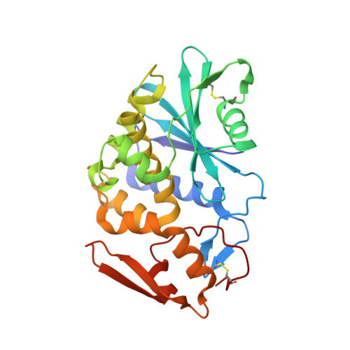
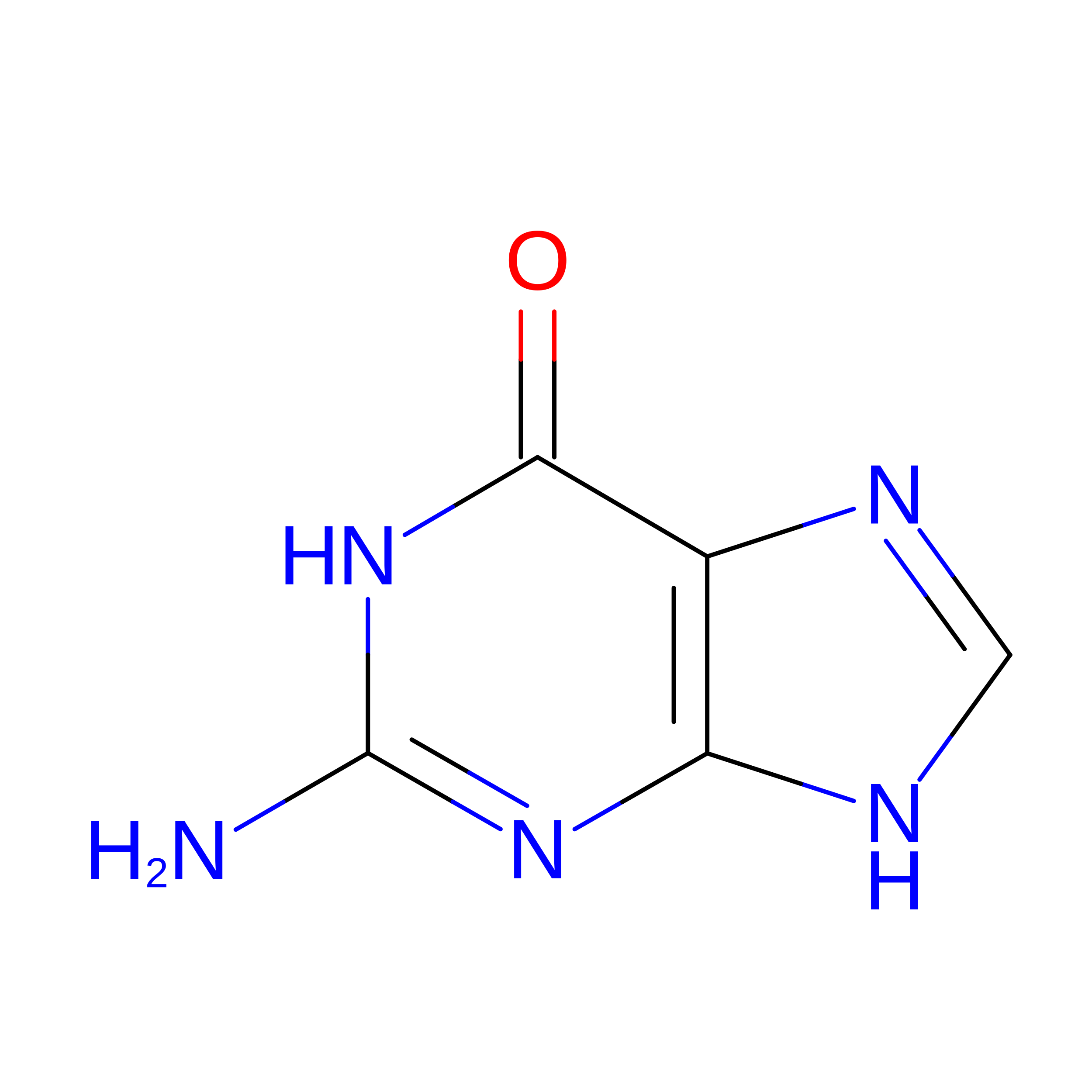
STRUCTURE OF POKEWEED ANTIVIRAL PROTEIN COMPLEXED WITH GUANINE


Experimental Data Snapshot

  • Method: X-RAY DIFFRACTION
  • Resolution: 2.10 Å
  • R-Value Free: 
    0.250 (Depositor) 
  • R-Value Work: 
    0.190 (Depositor) 
  • R-Value Observed: 
    0.192 (Depositor) 

Starting Model: experimental
View more details

wwPDB Validation 3D Report Full Report

Validation slider image for 1D6A

This is version 1.5 of the entry. See complete history. 

Literature

X-ray crystallographic analysis of the structural basis for the interaction of pokeweed antiviral protein with guanine residues of ribosomal RNA.

Kurinov, I.V., Rajamohan, F., Venkatachalam, T.K., Uckun, F.M.

(1999) Protein Sci 8: 2399-2405

  • DOI: https://doi.org/10.1110/ps.8.11.2399
  • Primary Citation Related Structures: 
    1D6A

  • PubMed Abstract: 

    Pokeweed antiviral protein (PAP) is a ribosome-inactivating protein (RIP), which enzymatically removes a single adenine base from a conserved, surface exposed loop sequence of ribosomal rRNA. We now present unprecedented experimental evidence that PAP can release not only adenine but guanine as well from Escherichia coli rRNA, albeit at a rate 20 times slower than for adenine. We also report X-ray structure analysis and supporting modeling studies for the interactions of PAP with guanine. Our modeling studies indicated that PAP can accommodate a guanine base in the active site pocket without large conformational changes. This prediction was experimentally confirmed, since a guanine base was visible in the active site pocket of the crystal structure of the PAP-guanine complex.


  • Organizational Affiliation: 
    • Department of Structural Biology, Hughes Institute, Roseville, Minnesota 55113, USA. igor@ih.org

Macromolecule Content 

  • Total Structure Weight: 58.99 kDa 
  • Atom Count: 4,694 
  • Modeled Residue Count: 524 
  • Deposited Residue Count: 524 
  • Unique protein chains: 1

Macromolecules

Find similar proteins by:|  3D Structure
Entity ID: 1
MoleculeChains  Sequence LengthOrganismDetailsImage
POKEWEED ANTIVIRAL PROTEIN
A, B
262Phytolacca americanaMutation(s): 0 
EC: 3.2.2.22
UniProt
Find proteins for P10297 (Phytolacca americana)
Explore P10297 
Go to UniProtKB:  P10297
Entity Groups
Sequence Clusters30% Identity50% Identity70% Identity90% Identity95% Identity100% Identity
UniProt GroupP10297
Sequence Annotations
Expand
Reference Sequence

Small Molecules

Experimental Data & Validation

Experimental Data

  • Method: X-RAY DIFFRACTION
  • Resolution: 2.10 Å
  • R-Value Free:  0.250 (Depositor) 
  • R-Value Work:  0.190 (Depositor) 
  • R-Value Observed: 0.192 (Depositor) 
Space Group: P 1
Unit Cell:
Length ( Å )Angle ( ˚ )
a = 49.294α = 68.664
b = 49.411β = 81.036
c = 64.288γ = 63.87
Software Package:
Software NamePurpose
SCALEPACKdata scaling
X-PLORmodel building
X-PLORrefinement
X-PLORphasing

Structure Validation

View Full Validation Report


Entry History 

Deposition Data

Revision History  (Full details and data files)

  • Version 1.0: 1999-12-16
    Type: Initial release
  • Version 1.1: 2008-04-27
    Changes: Version format compliance
  • Version 1.2: 2011-07-13
    Changes: Version format compliance
  • Version 1.3: 2017-10-04
    Changes: Refinement description
  • Version 1.4: 2023-08-09
    Changes: Data collection, Database references, Derived calculations, Refinement description
  • Version 1.5: 2024-11-20
    Changes: Structure summary