1D06 | pdb_00001d06

STRUCTURAL BASIS OF DIMERIZATION AND SENSORY MECHANISMS OF OXYGEN-SENSING DOMAIN OF RHIZOBIUM MELILOTI FIXL DETERMINED AT 1.4A RESOLUTION


Experimental Data Snapshot

  • Method: X-RAY DIFFRACTION
  • Resolution: 1.40 Å
  • R-Value Free: 
    0.275 (Depositor) 
  • R-Value Work: 
    0.224 (Depositor) 

wwPDB Validation 3D Report Full Report

Validation slider image for 1D06

This is version 1.5 of the entry. See complete history

Literature

Sensory mechanism of oxygen sensor FixL from Rhizobium meliloti: crystallographic, mutagenesis and resonance Raman spectroscopic studies

Miyatake, H.Mukai, M.Park, S.-Y.Adachi, S.Tamura, K.Nakamura, H.Nakamura, K.Tsuchiya, T.Iizuka, T.Shiro, Y.

(2000) J Mol Biology 301: 415-431

  • DOI: https://doi.org/10.1006/jmbi.2000.3954
  • Primary Citation Related Structures: 
    1D06, 1EW0

  • PubMed Abstract: 

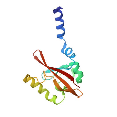
    FixL of Rhizobium meliloti (RmFixL) is a sensor histidine kinase of the two-component system, which regulates the expression of the genes related to nitrogen fixation in the root nodule in response to the O(2) levels. The crystal structure of the sensor domain of FixL (RmFixLH), which contains a heme (Fe-porphyrin) as a sensing site, was determined at 1.4 A resolution. Based on the structural and spectroscopic analyses, we propose the O(2) sensing mechanism that differs from the case proposed in BjFixLH as follows; conformational changes in the F/G loop, which are induced by steric repulsion between the bent-bound O(2) and the Ile209 side-chain, would be transmitted to the histidine kinase domain. Interaction between the iron-bound O(2) and Ile209 was also observed in the resonance Raman spectra of RmFixLH as evidenced by the fact that the Fe-O(2) and Fe-CN stretching frequencies were shifted from 575 to 570 cm(-1) (Fe-O(2)), and 504 to 499 cm(-1), respectively, as the result of the replacement of Ile209 with an Ala residue. In the I209A mutant of RmFixL, the O(2) sensing activity was destroyed, thus confirming our proposed mechanism.


  • Organizational Affiliation
    • RIKEN Harima Institute/SPring-8, 1-1-1 Koto, Hyogo, Mikazuki-cho, 679-5148, Japan. miyatake@postman.riken.go.jp

Macromolecule Content 

  • Total Structure Weight: 15.14 kDa 
  • Atom Count: 1,135 
  • Modeled Residue Count: 130 
  • Deposited Residue Count: 130 
  • Unique protein chains: 1

Macromolecules

Find similar proteins by:|  3D Structure
Entity ID: 1
MoleculeChains  Sequence LengthOrganismDetailsImage
nitrogen fixation regulatory protein fixL130Sinorhizobium melilotiMutation(s): 6 
EC: 2.7.13.3
UniProt
Find proteins for P10955 (Rhizobium meliloti (strain 1021))
Explore P10955 
Go to UniProtKB:  P10955
Entity Groups
Sequence Clusters30% Identity50% Identity70% Identity90% Identity95% Identity100% Identity
UniProt GroupP10955
Sequence Annotations
Expand
Reference Sequence

Small Molecules

Ligands 1 Unique
IDChains Name / Formula / InChI Key2D Diagram3D Interactions
HEM

Query on HEM


Download:Ideal Coordinates CCD File
B [auth A]PROTOPORPHYRIN IX CONTAINING FE
C34 H32 Fe N4 O4
KABFMIBPWCXCRK-RGGAHWMASA-L

Experimental Data & Validation

Experimental Data

  • Method: X-RAY DIFFRACTION
  • Resolution: 1.40 Å
  • R-Value Free:  0.275 (Depositor) 
  • R-Value Work:  0.224 (Depositor) 
Space Group: C 1 2 1
Unit Cell:
Length ( Å )Angle ( ˚ )
a = 60.6α = 90
b = 37.15β = 115.43
c = 53.99γ = 90
Software Package:
Software NamePurpose
DENZOdata reduction
SCALEPACKdata scaling
SOLVEphasing
X-PLORrefinement

Structure Validation

View Full Validation Report


Entry History 

Deposition Data

Revision History  (Full details and data files)

  • Version 1.0: 2000-03-15
    Type: Initial release
  • Version 1.1: 2008-04-27
    Changes: Version format compliance
  • Version 1.2: 2011-07-13
    Changes: Derived calculations, Version format compliance
  • Version 1.3: 2018-01-31
    Changes: Experimental preparation
  • Version 1.4: 2021-11-03
    Changes: Database references, Derived calculations
  • Version 1.5: 2024-02-07
    Changes: Data collection