1CWV

CRYSTAL STRUCTURE OF INVASIN: A BACTERIAL INTEGRIN-BINDING PROTEIN


Experimental Data Snapshot

  • Method: X-RAY DIFFRACTION
  • Resolution: 2.30 Å
  • R-Value Free: 0.274 
  • R-Value Work: 0.224 
  • R-Value Observed: 0.224 

wwPDB Validation   3D Report Full Report


This is version 1.2 of the entry. See complete history


Literature

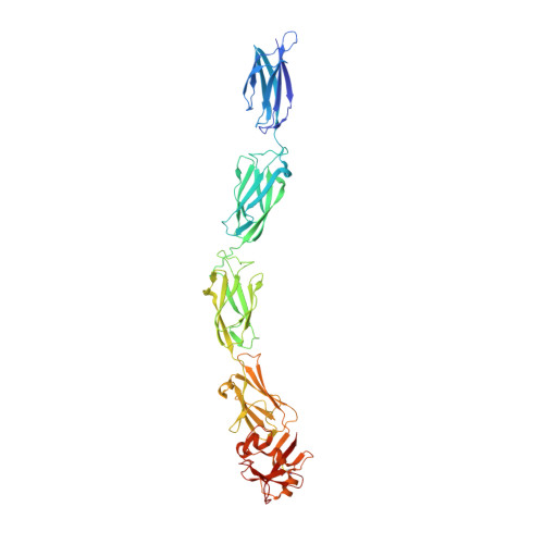
Crystal structure of invasin: a bacterial integrin-binding protein.

Hamburger, Z.A.Brown, M.S.Isberg, R.R.Bjorkman, P.J.

(1999) Science 286: 291-295

  • DOI: https://doi.org/10.1126/science.286.5438.291
  • Primary Citation of Related Structures:  
    1CWV

  • PubMed Abstract: 

    The Yersinia pseudotuberculosis invasin protein promotes bacterial entry by binding to host cell integrins with higher affinity than natural substrates such as fibronectin. The 2.3 angstrom crystal structure of the invasin extracellular region reveals five domains that form a 180 angstrom rod with structural similarities to tandem fibronectin type III domains. The integrin-binding surfaces of invasin and fibronectin include similarly located key residues, but in the context of different folds and surface shapes. The structures of invasin and fibronectin provide an example of convergent evolution, in which invasin presents an optimized surface for integrin binding, in comparison with host substrates.


  • Organizational Affiliation

    Division of Biology 156-29, Howard Hughes Medical Institute, California Institute of Technology, Pasadena, CA 91125, USA.


Macromolecules
Find similar proteins by:  (by identity cutoff)  |  3D Structure
Entity ID: 1
MoleculeChains Sequence LengthOrganismDetailsImage
INVASIN492Yersinia pseudotuberculosisMutation(s): 0 
UniProt
Find proteins for P11922 (Yersinia pseudotuberculosis serotype I (strain IP32953))
Explore P11922 
Go to UniProtKB:  P11922
Entity Groups  
Sequence Clusters30% Identity50% Identity70% Identity90% Identity95% Identity100% Identity
UniProt GroupP11922
Sequence Annotations
Expand
  • Reference Sequence
Small Molecules
Ligands 1 Unique
IDChains Name / Formula / InChI Key2D Diagram3D Interactions
CIT
Query on CIT

Download Ideal Coordinates CCD File 
B [auth A]CITRIC ACID
C6 H8 O7
KRKNYBCHXYNGOX-UHFFFAOYSA-N
Experimental Data & Validation

Experimental Data

  • Method: X-RAY DIFFRACTION
  • Resolution: 2.30 Å
  • R-Value Free: 0.274 
  • R-Value Work: 0.224 
  • R-Value Observed: 0.224 
  • Space Group: P 1 21 1
Unit Cell:
Length ( Å )Angle ( ˚ )
a = 61.14α = 90
b = 50.66β = 98.34
c = 97.92γ = 90
Software Package:
Software NamePurpose
DENZOdata reduction
SCALEPACKdata scaling
SHARPphasing
X-PLORrefinement

Structure Validation

View Full Validation Report


Entry History 

Deposition Data

Revision History  (Full details and data files)

  • Version 1.0: 1999-10-13
    Type: Initial release
  • Version 1.1: 2008-04-27
    Changes: Version format compliance
  • Version 1.2: 2011-07-13
    Changes: Version format compliance