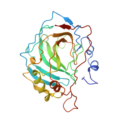
Structural consequences of redesigning a protein-zinc binding site.
Ippolito, J.A., Christianson, D.W.(1994) Biochemistry 33: 15241-15249
- PubMed: 7803386
- Primary Citation of Related Structures:
1CNB, 1CNC, 1CVD, 1CVE, 1CVF, 1CVH - PubMed Abstract:
In order to probe the structural importance of zinc ligands in the active site of human carbonic anhydrase II (CAII), we have determined the three-dimensional structures of H94C (in metal-bound form), H94C-BME (i.e., disulfide-linked with beta-mercaptoethanol), H94A, H96C, H119C, and H119D variants of CAII by X-ray crystallographic methods at resolutions of 2.2, 2.35, 2.25, 2.3, 2.2, and 2.25 A, respectively. Each variant crystallizes isomorphously with the wild-type enzyme, in which zinc is tetrahedrally coordinated by H94, H96, H119, and hydroxide ion. The structure of H94C CAII reveals the successful substitution of the naturally occurring histidine zinc ligand by a cysteine thiolate, and metal coordination by C94 is facilitated by the plastic structural response of the beta-sheet superstructure. Importantly, the resulting structure represents the catalytically active form of the enzyme reported previously [Alexander, R. S., Kiefer, L. L., Fierke, C. A., & Christianson, D. W. (1993) Biochemistry 32, 1510-1518]. Contrastingly, the structure of H96C CAII reveals that the engineered side chain does not coordinate to zinc; instead, zinc is tetrahedrally liganded by H94, H119, and two solvent molecules. Thus, the beta-sheet superstructure is not sufficiently plastic in this location to allow C96 to coordinate to the metal ion. Substitution of the thiolate or carboxylate group for wild-type histidine in H119C and H119D CAIIs reveals that tetrahedral metal coordination is maintained in each variant; however, since there is no plastic structural response of the corresponding beta-strand, a longer metal-ligand separation results.(ABSTRACT TRUNCATED AT 250 WORDS)
- Department of Chemistry, University of Pennsylvania, Philadelphia 19104-6323.
Organizational Affiliation:


