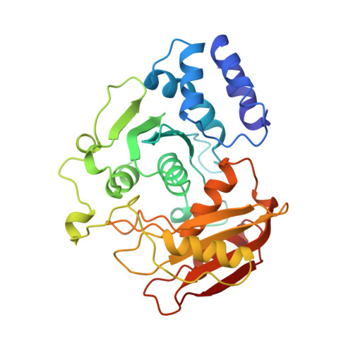
1CTU

TRANSITION-STATE SELECTIVITY FOR A SINGLE OH GROUP DURING CATALYSIS BY CYTIDINE DEAMINASE


Experimental Data Snapshot

  • Method: X-RAY DIFFRACTION
  • Resolution: 2.30 Å
  • R-Value Work: 0.190 
  • R-Value Observed: 0.190 

wwPDB Validation   3D Report Full Report


Ligand Structure Quality Assessment 


This is version 1.5 of the entry. See complete history


Literature

Transition-state selectivity for a single hydroxyl group during catalysis by cytidine deaminase.

Xiang, S.Short, S.A.Wolfenden, R.Carter Jr., C.W.

(1995) Biochemistry 34: 4516-4523

  • DOI: https://doi.org/10.1021/bi00014a003
  • Primary Citation of Related Structures:  
    1CTT, 1CTU

  • PubMed Abstract: 

    Cytidine deaminase binds transition-state analog inhibitors approximately 10(7) times more tightly than corresponding 3,4-dihydro analogs containing a proton in place of the 4-hydroxyl group. X-ray crystal structures of complexes with the two matched inhibitors differ only near a "trapped" water molecule in the complex with the 3,4-dihydro analog, where contacts are substantially less favorable than those with the hydroxyl group of the transition-state analog. The hydrogen bond between the hydroxyl group and the Glu 104 carboxylate shortens in that complex, and may become a "low-barrier" hydrogen bond, since at the same time the bond between zinc and the Cys 132 thiolate ligand lengthens. These differences must therefore account for most of the differential binding affinity related to catalysis. Moreover, the trapped water molecule retains some of the binding energy stabilizing the hydroxyl group in the transition-state analog complex. To this extent, the ratio of binding affinities for the two compounds is smaller than the true contribution of the hydroxyl group, a conclusion with significant bearing on interpreting difference free energies derived from substituent effects arising from chemical modification and/or mutagenesis.


  • Organizational Affiliation

    Department of Biochemistry and Biophysics, University of North Carolina at Chapel Hill 27599-7260, USA.


Macromolecules
Find similar proteins by:  (by identity cutoff)  |  3D Structure
Entity ID: 1
MoleculeChains Sequence LengthOrganismDetailsImage
CYTIDINE DEAMINASE294Escherichia coliMutation(s): 0 
EC: 3.5.4.5
UniProt
Find proteins for P0ABF6 (Escherichia coli (strain K12))
Explore P0ABF6 
Go to UniProtKB:  P0ABF6
Entity Groups  
Sequence Clusters30% Identity50% Identity70% Identity90% Identity95% Identity100% Identity
UniProt GroupP0ABF6
Sequence Annotations
Expand
  • Reference Sequence
Small Molecules
Binding Affinity Annotations 
IDSourceBinding Affinity
ZEB PDBBind:  1CTU Ki: 1.20e-3 (nM) from 1 assay(s)
Experimental Data & Validation

Experimental Data

  • Method: X-RAY DIFFRACTION
  • Resolution: 2.30 Å
  • R-Value Work: 0.190 
  • R-Value Observed: 0.190 
  • Space Group: P 31 2 1
Unit Cell:
Length ( Å )Angle ( ˚ )
a = 120.3α = 90
b = 120.3β = 90
c = 78.4γ = 120
Software Package:
Software NamePurpose
X-PLORmodel building
TNTrefinement
X-PLORrefinement
R-AXISdata reduction
X-PLORphasing

Structure Validation

View Full Validation Report


Ligand Structure Quality Assessment 


Entry History 

Deposition Data

Revision History  (Full details and data files)

  • Version 1.0: 1995-05-08
    Type: Initial release
  • Version 1.1: 2008-03-03
    Changes: Version format compliance
  • Version 1.2: 2011-07-13
    Changes: Derived calculations, Version format compliance
  • Version 1.3: 2019-07-17
    Changes: Data collection, Other, Refinement description
  • Version 1.4: 2019-08-14
    Changes: Data collection, Refinement description
  • Version 1.5: 2024-02-07
    Changes: Data collection, Database references, Derived calculations