1CR5 | pdb_00001cr5

N-TERMINAL DOMAIN OF SEC18P


Experimental Data Snapshot

  • Method: X-RAY DIFFRACTION
  • Resolution: 2.30 Å
  • R-Value Free: 
    0.277 (Depositor), 0.274 (DCC) 
  • R-Value Work: 
    0.230 (Depositor), 0.225 (DCC) 

wwPDB Validation 3D Report Full Report

Validation slider image for 1CR5

This is version 1.4 of the entry. See complete history

Literature

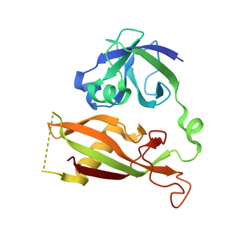
Crystal structure of the Sec18p N-terminal domain.

Babor, S.M.Fass, D.

(1999) Proc Natl Acad Sci U S A 96: 14759-14764

  • DOI: https://doi.org/10.1073/pnas.96.26.14759
  • Primary Citation Related Structures: 
    1CR5

  • PubMed Abstract: 

    Yeast Sec18p and its mammalian orthologue N-ethylmaleimide-sensitive fusion protein (NSF) are hexameric ATPases with a central role in vesicle trafficking. Aided by soluble adapter factors (SNAPs), Sec18p/NSF induces ATP-dependent disassembly of a complex of integral membrane proteins from the vesicle and target membranes (SNAP receptors). During the ATP hydrolysis cycle, the Sec18p/NSF homohexamer undergoes a large-scale conformational change involving repositioning of the most N terminal of the three domains of each protomer, a domain that is required for SNAP-mediated interaction with SNAP receptors. Whether an internal conformational change in the N-terminal domains accompanies their reorientation with respect to the rest of the hexamer remains to be addressed. We have determined the structure of the N-terminal domain from Sec18p by x-ray crystallography. The Sec18p N-terminal domain consists of two beta-sheet-rich subdomains connected by a short linker. A conserved basic cleft opposite the linker may constitute a SNAP-binding site. Despite structural variability in the linker region and in an adjacent loop, all three independent molecules in the crystal asymmetric unit have the identical subdomain interface, supporting the notion that this interface is a preferred packing arrangement. However, the linker flexibility allows for the possibility that other subdomain orientations may be sampled.


  • Organizational Affiliation
    • Department of Structural Biology, Weizmann Institute of Science, Rehovot 76100, Israel.

Macromolecule Content 

  • Total Structure Weight: 64.73 kDa 
  • Atom Count: 4,555 
  • Modeled Residue Count: 529 
  • Deposited Residue Count: 567 
  • Unique protein chains: 1

Macromolecules

Find similar proteins by:|  3D Structure
Entity ID: 1
MoleculeChains  Sequence LengthOrganismDetailsImage
SEC18P (RESIDUES 22 - 210)
A, B, C
189Saccharomyces cerevisiaeMutation(s): 0 
UniProt
Find proteins for P18759 (Saccharomyces cerevisiae (strain ATCC 204508 / S288c))
Explore P18759 
Go to UniProtKB:  P18759
Entity Groups
Sequence Clusters30% Identity50% Identity70% Identity90% Identity95% Identity100% Identity
UniProt GroupP18759
Sequence Annotations
Expand
Reference Sequence

Experimental Data & Validation

Experimental Data

  • Method: X-RAY DIFFRACTION
  • Resolution: 2.30 Å
  • R-Value Free:  0.277 (Depositor), 0.274 (DCC) 
  • R-Value Work:  0.230 (Depositor), 0.225 (DCC) 
Space Group: P 1 21 1
Unit Cell:
Length ( Å )Angle ( ˚ )
a = 93.71α = 90
b = 32.14β = 118.7
c = 94.85γ = 90
Software Package:
Software NamePurpose
SHARPphasing
CNSrefinement
DENZOdata reduction
SCALEPACKdata scaling

Structure Validation

View Full Validation Report


Entry History 

Deposition Data

Revision History  (Full details and data files)

  • Version 1.0: 1999-12-22
    Type: Initial release
  • Version 1.1: 2008-04-27
    Changes: Version format compliance
  • Version 1.2: 2011-07-13
    Changes: Version format compliance
  • Version 1.3: 2018-01-31
    Changes: Experimental preparation
  • Version 1.4: 2024-10-16
    Changes: Data collection, Database references, Derived calculations, Structure summary