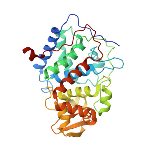
1CPG

A CATION BINDING MOTIF STABILIZES THE COMPOUND I RADICAL OF CYTOCHROME C PEROXIDASE


Experimental Data Snapshot

  • Method: X-RAY DIFFRACTION
  • Resolution: 2.20 Å
  • R-Value Work: 0.164 
  • R-Value Observed: 0.164 

wwPDB Validation   3D Report Full Report


Ligand Structure Quality Assessment 


This is version 1.6 of the entry. See complete history


Literature

A cation binding motif stabilizes the compound I radical of cytochrome c peroxidase.

Miller, M.A.Han, G.W.Kraut, J.

(1994) Proc Natl Acad Sci U S A 91: 11118-11122

  • DOI: https://doi.org/10.1073/pnas.91.23.11118
  • Primary Citation of Related Structures:  
    1CPD, 1CPE, 1CPF, 1CPG

  • PubMed Abstract: 

    Cytochrome c peroxidase reacts with peroxide to form compound I, which contains an oxyferryl heme and an indolyl radical at Trp-191. The indolyl free radical has a half-life of several hours at room temperature, and this remarkable stability is essential for the catalytic function of cytochrome c peroxidase. To probe the protein environment that stabilizes the compound I radical, we used site-directed mutagenesis to replace Trp-191 with Gly or Gln. Crystal structures of these mutants revealed a monovalent cation binding site in the cavity formerly occupied by the side chain of Trp-191. Comparison of this site with those found in other known cation binding enzymes shows that the Trp-191 side chain resides in a consensus K+ binding site. Electrostatic potential calculations indicate that the cation binding site is created by partial negative charges at the backbone carbonyl oxygen atoms of residues 175 and 177, the carboxyl end of a long alpha-helix (residues 165-175), the heme propionates, and the carboxylate side chain of Asp-235. These features create a negative potential that envelops the side chain of Trp-191; the calculated free energy change for cation binding in this site is -27 kcal/mol (1 cal = 4.184J). This is more than sufficient to account for the stability of the Trp-191 radical, which our estimates suggest is stabilized by 7.8 kcal/mol relative to a Trp radical in solution.


  • Organizational Affiliation

    Department of Chemistry, University of California at San Diego, La Jolla 92093-0317.


Macromolecules
Find similar proteins by:  (by identity cutoff)  |  3D Structure
Entity ID: 1
MoleculeChains Sequence LengthOrganismDetailsImage
CYTOCHROME C PEROXIDASE296Saccharomyces cerevisiaeMutation(s): 0 
EC: 1.11.1.5
UniProt
Find proteins for P00431 (Saccharomyces cerevisiae (strain ATCC 204508 / S288c))
Explore P00431 
Go to UniProtKB:  P00431
Entity Groups  
Sequence Clusters30% Identity50% Identity70% Identity90% Identity95% Identity100% Identity
UniProt GroupP00431
Sequence Annotations
Expand
  • Reference Sequence
Experimental Data & Validation

Experimental Data

  • Method: X-RAY DIFFRACTION
  • Resolution: 2.20 Å
  • R-Value Work: 0.164 
  • R-Value Observed: 0.164 
  • Space Group: P 21 21 21
Unit Cell:
Length ( Å )Angle ( ˚ )
a = 105.17α = 90
b = 74.18β = 90
c = 45.22γ = 90
Software Package:
Software NamePurpose
X-PLORmodel building
TNTrefinement
X-PLORrefinement
X-PLORphasing

Structure Validation

View Full Validation Report


Ligand Structure Quality Assessment 


Entry History 

Deposition Data

Revision History  (Full details and data files)

  • Version 1.0: 1994-11-01
    Type: Initial release
  • Version 1.1: 2008-03-24
    Changes: Version format compliance
  • Version 1.2: 2011-07-13
    Changes: Version format compliance
  • Version 1.3: 2017-11-29
    Changes: Derived calculations, Other
  • Version 1.4: 2019-07-17
    Changes: Data collection, Refinement description
  • Version 1.5: 2019-08-14
    Changes: Data collection, Refinement description
  • Version 1.6: 2024-02-07
    Changes: Data collection, Database references, Derived calculations