1CPC | pdb_00001cpc

ISOLATION, CRYSTALLIZATION, CRYSTAL STRUCTURE ANALYSIS AND REFINEMENT OF CONSTITUTIVE C-PHYCOCYANIN FROM THE CHROMATICALLY ADAPTING CYANOBACTERIUM FREMYELLA DIPLOSIPHON AT 1.66 ANGSTROMS RESOLUTION


Experimental Data Snapshot

  • Method: X-RAY DIFFRACTION
  • Resolution: 1.66 Å
  • R-Value Work: 
    0.181 (Depositor) 

wwPDB Validation 3D Report Full Report

Validation slider image for 1CPC

This is version 1.4 of the entry. See complete history

Literature

Isolation, crystallization, crystal structure analysis and refinement of constitutive C-phycocyanin from the chromatically adapting cyanobacterium Fremyella diplosiphon at 1.66 A resolution.

Duerring, M.Schmidt, G.B.Huber, R.

(1991) J Mol Biology 217: 577-592

  • DOI: https://doi.org/10.1016/0022-2836(91)90759-y
  • Primary Citation Related Structures: 
    1CPC

  • PubMed Abstract: 

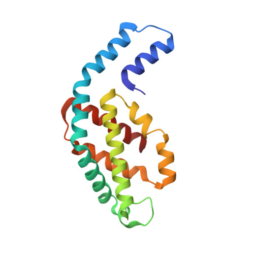
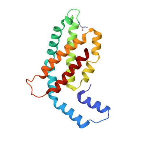
    Constitutive phycocyanin from cyanobacterium Fremyella diplosiphon (Calothrix sp. PCC 7601) grown in green light, has been isolated and crystallized. The crystals belong to the space group R3 with cell constants a = b = 180.26 A, c = 61.24 A, alpha = beta = 90 degrees, gamma = 120 degrees. The crystal structure has been determined by Patterson search techniques using the molecular model of C-phycocyanin from the cyanobacterium Agmenellum quadruplicatum. The asymmetric unit of the crystal cell consists of two (alpha beta)-monomers related by a local dyad. Three asymmetric units are arranged around a crystallographic triad and form an (alpha beta)6-hexamer, the functional unit in the native antenna rod. The initial structure has been refined in a cyclic manner by energy-restrained crystallographic refinement and modelling until the conventional crystallographic R-factor converged at 18.1% with data to a resolution of 1.66 A. The molecular structure resembles closely the C-phycocyanins of Mastigocladus laminosus and A. quadruplicatum. The conformation and configuration of the alpha-84 and beta-84 chromophores is very similar to the corresponding chromophores in the trimeric C-phycocyanin of M. laminosus, whereas the beta-155 chromophore differs in configuration with C(4)-Z, C(10)-Z and C(15)-Z compared to C(4)-Z, C(10)-Z, C(15)-Z,E. The stereochemistry of the beta-155 chiral centres is C(2)-RC(3)-R and C(31)-S, respectively, whereas alpha-84 and beta-84 have C(2)-RC(3)-R and C(31)-R. The amino acid sequences of constitutive and inducible phycocyanin differ mainly in residues located on the surface of the beta-subunits that mediate the inter-hexameric contacts.


  • Organizational Affiliation
    • Max-Planck-Institut für Biochemie, München, Germany.

Macromolecule Content 

  • Total Structure Weight: 73.92 kDa 
  • Atom Count: 5,434 
  • Modeled Residue Count: 668 
  • Deposited Residue Count: 668 
  • Unique protein chains: 2

Macromolecules

Find similar proteins by:|  3D Structure
Entity ID: 1
MoleculeChains  Sequence LengthOrganismDetailsImage
C-PHYCOCYANIN (ALPHA SUBUNIT)A,
C [auth K]
162Microchaete diplosiphonMutation(s): 0 
UniProt
Find proteins for P07122 (Microchaete diplosiphon)
Explore P07122 
Go to UniProtKB:  P07122
Entity Groups
Sequence Clusters30% Identity50% Identity70% Identity90% Identity95% Identity100% Identity
UniProt GroupP07122
Sequence Annotations
Expand
Reference Sequence
Find similar proteins by:|  3D Structure
Entity ID: 2
MoleculeChains  Sequence LengthOrganismDetailsImage
C-PHYCOCYANIN (BETA SUBUNIT)B,
D [auth L]
172Microchaete diplosiphonMutation(s): 0 
UniProt
Find proteins for P07119 (Microchaete diplosiphon)
Explore P07119 
Go to UniProtKB:  P07119
Entity Groups
Sequence Clusters30% Identity50% Identity70% Identity90% Identity95% Identity100% Identity
UniProt GroupP07119
Sequence Annotations
Expand
Reference Sequence

Experimental Data & Validation

Experimental Data

  • Method: X-RAY DIFFRACTION
  • Resolution: 1.66 Å
  • R-Value Work:  0.181 (Depositor) 
Space Group: H 3
Unit Cell:
Length ( Å )Angle ( ˚ )
a = 180.26α = 90
b = 180.26β = 90
c = 61.24γ = 120
Software Package:
Software NamePurpose
EREFrefinement

Structure Validation

View Full Validation Report


Entry History 

Deposition Data

Revision History  (Full details and data files)

  • Version 1.0: 1993-01-15
    Type: Initial release
  • Version 1.1: 2008-03-03
    Changes: Version format compliance
  • Version 1.2: 2011-07-13
    Changes: Version format compliance
  • Version 1.3: 2013-04-10
    Changes: Non-polymer description, Structure summary
  • Version 1.4: 2024-06-05
    Changes: Data collection, Database references, Derived calculations, Other