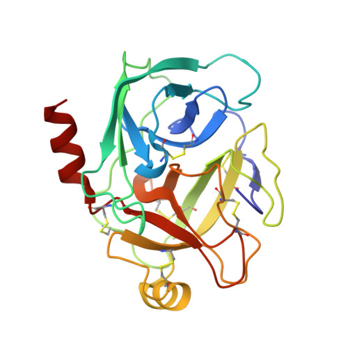
1C5Q

STRUCTURAL BASIS FOR SELECTIVITY OF A SMALL MOLECULE, S1-BINDING, SUB-MICROMOLAR INHIBITOR OF UROKINASE TYPE PLASMINOGEN ACTIVATOR


Experimental Data Snapshot

  • Method: X-RAY DIFFRACTION
  • Resolution: 1.43 Å
  • R-Value Free: 0.221 
  • R-Value Work: 0.197 
  • R-Value Observed: 0.197 

wwPDB Validation   3D Report Full Report


Ligand Structure Quality Assessment 


This is version 1.5 of the entry. See complete history


Literature

Structural basis for selectivity of a small molecule, S1-binding, submicromolar inhibitor of urokinase-type plasminogen activator.

Katz, B.A.Mackman, R.Luong, C.Radika, K.Martelli, A.Sprengeler, P.A.Wang, J.Chan, H.Wong, L.

(2000) Chem Biol 7: 299-312

  • DOI: https://doi.org/10.1016/s1074-5521(00)00104-6
  • Primary Citation of Related Structures:  
    1C5L, 1C5M, 1C5N, 1C5O, 1C5P, 1C5Q, 1C5R, 1C5S, 1C5T, 1C5U, 1C5V, 1C5W, 1C5X, 1C5Y, 1C5Z

  • PubMed Abstract: 

    Urokinase-type plasminogen activator (uPA) is a protease associated with tumor metastasis and invasion. Inhibitors of uPA may have potential as drugs for prostate, breast and other cancers. Therapeutically useful inhibitors must be selective for uPA and not appreciably inhibit the related, and structurally and functionally similar enzyme, tissue-type plasminogen activator (tPA), involved in the vital blood-clotting cascade. We produced mutagenically deglycosylated low molecular weight uPA and determined the crystal structure of its complex with 4-iodobenzo[b]thiophene 2-carboxamidine (K(i) = 0.21 +/- 0.02 microM). To probe the structural determinants of the affinity and selectivity of this inhibitor for uPA we also determined the structures of its trypsin and thrombin complexes, of apo-trypsin, apo-thrombin and apo-factor Xa, and of uPA, trypsin and thrombin bound by compounds that are less effective uPA inhibitors, benzo[b]thiophene-2-carboxamidine, thieno[2,3-b]-pyridine-2-carboxamidine and benzamidine. The K(i) values of each inhibitor toward uPA, tPA, trypsin, tryptase, thrombin and factor Xa were determined and compared. One selectivity determinant of the benzo[b]thiophene-2-carboxamidines for uPA involves a hydrogen bond at the S1 site to Ogamma(Ser190) that is absent in the Ala190 proteases, tPA, thrombin and factor Xa. Other subtle differences in the architecture of the S1 site also influence inhibitor affinity and enzyme-bound structure. Subtle structural differences in the S1 site of uPA compared with that of related proteases, which result in part from the presence of a serine residue at position 190, account for the selectivity of small thiophene-2-carboxamidines for uPA, and afford a framework for structure-based design of small, potent, selective uPA inhibitors.


  • Organizational Affiliation

    Axys Pharmaceutical Corporation, South San Francisco, CA 94080, USA. brad_katz@axyspharm.com


Macromolecules
Find similar proteins by:  (by identity cutoff)  |  3D Structure
Entity ID: 1
MoleculeChains Sequence LengthOrganismDetailsImage
PROTEIN (TRYPSIN)223Bos taurusMutation(s): 0 
EC: 3.4.21.4
UniProt
Find proteins for P00760 (Bos taurus)
Explore P00760 
Go to UniProtKB:  P00760
Entity Groups  
Sequence Clusters30% Identity50% Identity70% Identity90% Identity95% Identity100% Identity
UniProt GroupP00760
Sequence Annotations
Expand
  • Reference Sequence
Small Molecules
Ligands 3 Unique
IDChains Name / Formula / InChI Key2D Diagram3D Interactions
ESI
Query on ESI

Download Ideal Coordinates CCD File 
D [auth A]4-IODOBENZO[B]THIOPHENE-2-CARBOXAMIDINE
C9 H8 I N2 S
YERQOXAYAFWFEJ-UHFFFAOYSA-O
CA
Query on CA

Download Ideal Coordinates CCD File 
B [auth A]CALCIUM ION
Ca
BHPQYMZQTOCNFJ-UHFFFAOYSA-N
CL
Query on CL

Download Ideal Coordinates CCD File 
C [auth A]CHLORIDE ION
Cl
VEXZGXHMUGYJMC-UHFFFAOYSA-M
Binding Affinity Annotations 
IDSourceBinding Affinity
ESI PDBBind:  1C5Q Ki: 440 (nM) from 1 assay(s)
Experimental Data & Validation

Experimental Data

  • Method: X-RAY DIFFRACTION
  • Resolution: 1.43 Å
  • R-Value Free: 0.221 
  • R-Value Work: 0.197 
  • R-Value Observed: 0.197 
  • Space Group: P 21 21 21
Unit Cell:
Length ( Å )Angle ( ˚ )
a = 63.87α = 90
b = 64.11β = 90
c = 69.49γ = 90
Software Package:
Software NamePurpose
bioteXdata collection
bioteXdata reduction
X-PLORmodel building
Quantamodel building
Insight IImodel building
X-PLORrefinement
bioteXdata scaling
X-PLORphasing

Structure Validation

View Full Validation Report


Ligand Structure Quality Assessment 


Entry History 

Deposition Data

Revision History  (Full details and data files)

  • Version 1.0: 2000-12-22
    Type: Initial release
  • Version 1.1: 2008-04-26
    Changes: Version format compliance
  • Version 1.2: 2011-07-13
    Changes: Version format compliance
  • Version 1.3: 2017-10-04
    Changes: Advisory, Refinement description
  • Version 1.4: 2023-12-27
    Changes: Advisory, Data collection, Database references, Derived calculations
  • Version 1.5: 2024-10-16
    Changes: Structure summary