1BQT | pdb_00001bqt

THREE-DIMENSIONAL STRUCTURE OF HUMAN INSULIN-LIKE GROWTH FACTOR-I (IGF-I) DETERMINED BY 1H-NMR AND DISTANCE GEOMETRY, 6 STRUCTURES


Experimental Data Snapshot

  • Method: SOLUTION NMR
  • Conformers Calculated: 100 
  • Conformers Submitted: 
  • Selection Criteria: target function 

wwPDB Validation 3D Report Full Report

Validation slider image for 1BQT

This is version 1.4 of the entry. See complete history

Literature

Three-dimensional structure of human insulin-like growth factor-I (IGF-I) determined by 1H-NMR and distance geometry.

Sato, A.Nishimura, S.Ohkubo, T.Kyogoku, Y.Koyama, S.Kobayashi, M.Yasuda, T.Kobayashi, Y.

(1993) Int J Pept Protein Res 41: 433-440

  • DOI: https://doi.org/10.1111/j.1399-3011.1993.tb00462.x
  • Primary Citation Related Structures: 
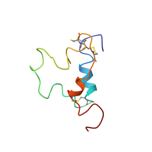
    1BQT

  • PubMed Abstract: 

    The three-dimensional structure of human insulin-like growth factor-I has been determined through a combination of NMR measurements and distance geometry calculations. A total of 320 interatomic distance constraints, including 12 related to the disulfide bridges, were used in these calculations. The resulting structure is characterized by the presence of three helical rods corresponding to the sequence regions, Ala8-Cys18, Gly42-Cys48 and Leu54-Cys61. Furthermore, a turn structure and an extended structure exist in the Gly19-Gly22 and Phe23-Asn26 regions, respectively. Neglecting the N- and C-termini, with their expectedly high degree of mobility as well as a fluctuating C-domain, the r.m.s.d. value is 1.9 A for backbone atoms. Those of the three alpha-helical regions are 1.0, 0.9 and 0.8 A, respectively, 1.8 A being that for the total backbone atoms participating in the formation of these three helices, showing the good convergence of their spatial arrangements. The overall structure obtained here shows that the human IGF-I molecule folds into a spatial structure very similar to that of insulin in an aqueous solution.


  • Organizational Affiliation
    • Institute for Protein Research, Osaka University, Japan.

Macromolecule Content 

  • Total Structure Weight: 7.66 kDa 
  • Atom Count: 533 
  • Modeled Residue Count: 70 
  • Deposited Residue Count: 70 
  • Unique protein chains: 1

Macromolecules

Find similar proteins by:|  3D Structure
Entity ID: 1
MoleculeChains  Sequence LengthOrganismDetailsImage
INSULIN-LIKE GROWTH FACTOR-I70Homo sapiensMutation(s): 0 
UniProt & NIH Common Fund Data Resources
Find proteins for P05019 (Homo sapiens)
Explore P05019 
Go to UniProtKB:  P05019
PHAROS:  P05019
GTEx:  ENSG00000017427 
Entity Groups
Sequence Clusters30% Identity50% Identity70% Identity90% Identity95% Identity100% Identity
UniProt GroupP05019
Sequence Annotations
Expand
Reference Sequence

Experimental Data & Validation

Experimental Data

  • Method: SOLUTION NMR
  • Conformers Calculated: 100 
  • Conformers Submitted: 
  • Selection Criteria: target function 

Structure Validation

View Full Validation Report


Entry History 

Deposition Data

Revision History  (Full details and data files)

  • Version 1.0: 1999-05-18
    Type: Initial release
  • Version 1.1: 2008-03-24
    Changes: Version format compliance
  • Version 1.2: 2011-07-13
    Changes: Version format compliance
  • Version 1.3: 2022-02-16
    Changes: Database references, Derived calculations, Other
  • Version 1.4: 2024-10-30
    Changes: Data collection, Structure summary