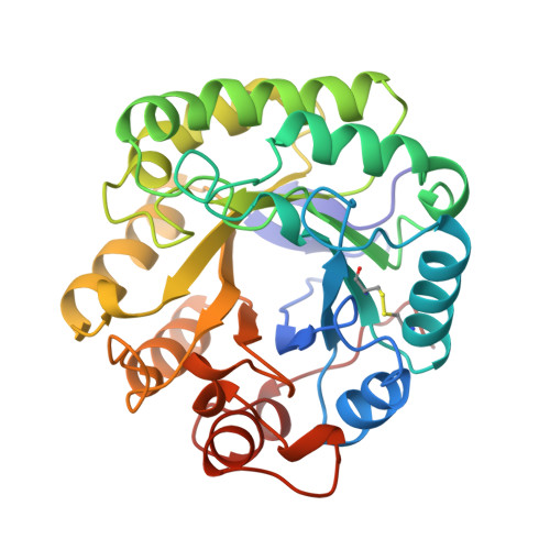
High-resolution native and complex structures of thermostable beta-mannanase from Thermomonospora fusca - substrate specificity in glycosyl hydrolase family 5.
Hilge, M., Gloor, S.M., Rypniewski, W., Sauer, O., Heightman, T.D., Zimmermann, W., Winterhalter, K., Piontek, K.(1998) Structure 6: 1433-1444
- PubMed: 9817845 Search on PubMed
- DOI: https://doi.org/10.1016/s0969-2126(98)00142-7
- Primary Citation Related Structures:
1BQC, 2MAN, 3MAN - PubMed Abstract:
. beta-Mannanases hydrolyse the O-glycosidic bonds in mannan, a hemicellulose constituent of plants. These enzymes have potential use in pulp and paper production and are of significant biotechnological interest. Thermostable beta-mannanases would be particularly useful due to their high temperature optimum and broad pH tolerance. The thermophilic actinomycete Thermomonospora fusca secretes at least one beta-mannanase (molecular mass 38 kDa) with a temperature optimum of 80 degreesC. No three-dimensional structure of a mannan-degrading enzyme has been reported until now. . The crystal structure of the thermostable beta-mannanase from T. fusca has been determined by the multiple isomorphous replacement method and refined to 1.5 A resolution. In addition to the native enzyme, the structures of the mannotriose- and mannohexaose-bound forms of the enzyme have been determined to resolutions of 1.9 A and 1.6 A, respectively. . Analysis of the -1 subsite of T. fusca mannanase reveals neither a favourable interaction towards the axial HO-C(2) nor a discrimination against the equatorial hydroxyl group of gluco-configurated substrates. We propose that selectivity arises from two possible mechanisms: a hydrophobic interaction of the substrate with Val263, conserved in family 5 bacterial mannanases, which discriminates between the different conformations of the hydroxymethyl group in native mannan and cellulose; and/or a specific interaction between Asp259 and the axial hydroxyl group at the C(2) of the substrate in the -2 subsite. Compared with the catalytic clefts of family 5 cellulases, the groove of T. fusca mannanase has a strongly reduced number of aromatic residues providing platforms for stacking with the substrate. This deletion of every second platform is in good agreement with the orientation of the axial hydroxyl groups in mannan.
- Laboratorium für Biochemie ETH Zentrum Universitätstrasse 16 CH-8092 Zürich, Switzerland.
Organizational Affiliation: