1B93 | pdb_00001b93

METHYLGLYOXAL SYNTHASE FROM ESCHERICHIA COLI


Experimental Data Snapshot

  • Method: X-RAY DIFFRACTION
  • Resolution: 1.90 Å
  • R-Value Free: 
    0.202 (Depositor), 0.189 (DCC) 
  • R-Value Work: 
    0.181 (Depositor), 0.172 (DCC) 
  • R-Value Observed: 
    0.181 (Depositor) 

wwPDB Validation 3D Report Full Report

Validation slider image for 1B93

This is version 1.3 of the entry. See complete history

Literature

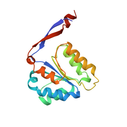
The crystal structure of methylglyoxal synthase from Escherichia coli.

Saadat, D.Harrison, D.H.

(1999) Structure 7: 309-317

  • DOI: https://doi.org/10.1016/s0969-2126(99)80041-0
  • Primary Citation Related Structures: 
    1B93

  • PubMed Abstract: 

    The reaction mechanism of methylglyoxal synthase (MGS) is believed to be similar to that of triosephosphate isomerase (TIM). Both enzymes utilise dihydroxyacetone phosphate (DHAP) to form an enediol(ate) phosphate intermediate as the first step of their reaction pathways. However, the second catalytic step in the MGS reaction pathway is characterized by the elimination of phosphate and collapse of the enediol(ate) to form methylglyoxal instead of reprotonation to form the isomer glyceraldehyde 3-phosphate. The crystal structure of MGS bound to formate and substoichiometric amounts of phosphate in the space group P6522 has been determined at 1.9 A resolution. This structure shows that the enzyme is a homohexamer composed of interacting five-stranded beta/alpha proteins, rather than the hallmark alpha/beta barrel structure of TIM. The conserved residues His19, Asp71, and His98 in each of the three monomers in the asymmetric unit bind to a formate ion that is present in the crystallization conditions. Differences in the three monomers in the asymmetric unit are localized at the mouth of the active site and can be ascribed to the presence or absence of a bound phosphate ion. In agreement with site-directed mutagenesis and mechanistic enzymology, the structure suggests that Asp71 acts as the catalytic base. Further, Asp20 and Asp101 are involved in intersubunit salt bridges. These salt bridges may provide a pathway for transmitting allosteric information.


  • Organizational Affiliation
    • Department of Biochemistry, Medical College of Wisconsin, 8701 Watertown Plank Road, Milwaukee, WI 53226, USA.

Macromolecule Content 

  • Total Structure Weight: 51.23 kDa 
  • Atom Count: 3,639 
  • Modeled Residue Count: 443 
  • Deposited Residue Count: 456 
  • Unique protein chains: 1

Macromolecules

Find similar proteins by:|  3D Structure
Entity ID: 1
MoleculeChains  Sequence LengthOrganismDetailsImage
PROTEIN (METHYLGLYOXAL SYNTHASE)
A, B, C
152Escherichia coliMutation(s): 0 
Gene Names: MGSA
EC: 4.2.3.3
UniProt
Find proteins for P0A731 (Escherichia coli (strain K12))
Explore P0A731 
Go to UniProtKB:  P0A731
Entity Groups
Sequence Clusters30% Identity50% Identity70% Identity90% Identity95% Identity100% Identity
UniProt GroupP0A731
Sequence Annotations
Expand
Reference Sequence

Small Molecules

Ligands 2 Unique
IDChains Name / Formula / InChI Key2D Diagram3D Interactions
PO4

Query on PO4


Download:Ideal Coordinates CCD File
G [auth B]PHOSPHATE ION
O4 P
NBIIXXVUZAFLBC-UHFFFAOYSA-K
FMT

Query on FMT


Download:Ideal Coordinates CCD File
D [auth A]
E [auth A]
F [auth A]
H [auth B]
I [auth C]
D [auth A],
E [auth A],
F [auth A],
H [auth B],
I [auth C],
J [auth C],
K [auth C]
FORMIC ACID
C H2 O2
BDAGIHXWWSANSR-UHFFFAOYSA-N

Experimental Data & Validation

Experimental Data

  • Method: X-RAY DIFFRACTION
  • Resolution: 1.90 Å
  • R-Value Free:  0.202 (Depositor), 0.189 (DCC) 
  • R-Value Work:  0.181 (Depositor), 0.172 (DCC) 
  • R-Value Observed: 0.181 (Depositor) 
Space Group: P 65 2 2
Unit Cell:
Length ( Å )Angle ( ˚ )
a = 123.34α = 90
b = 123.34β = 90
c = 155.98γ = 120
Software Package:
Software NamePurpose
XTALVIEWrefinement
X-PLORrefinement
DENZOdata reduction
SCALEPACKdata scaling

Structure Validation

View Full Validation Report


Entry History 

Deposition Data

Revision History  (Full details and data files)

  • Version 1.0: 1999-03-16
    Type: Initial release
  • Version 1.1: 2008-04-26
    Changes: Version format compliance
  • Version 1.2: 2011-07-13
    Changes: Derived calculations, Version format compliance
  • Version 1.3: 2023-12-27
    Changes: Data collection, Database references, Derived calculations