1B8X | pdb_00001b8x

GLUTATHIONE S-TRANSFERASE FUSED WITH THE NUCLEAR MATRIX TARGETING SIGNAL OF THE TRANSCRIPTION FACTOR AML-1


Experimental Data Snapshot

  • Method: X-RAY DIFFRACTION
  • Resolution: 2.70 Å
  • R-Value Free: 
    0.310 (Depositor) 
  • R-Value Work: 
    0.209 (Depositor) 

Starting Model: experimental
View more details

wwPDB Validation 3D Report Full Report

Validation slider image for 1B8X

This is version 1.3 of the entry. See complete history

Literature

Preliminary crystallographic study of glutathione S-transferase fused with the nuclear matrix targeting signal of the transcription factor AML-1/CBF-alpha2.

Tang, L.Guo, B.van Wijnen, A.J.Lian, J.B.Stein, J.L.Stein, G.S.Zhou, G.W.

(1998) J Struct Biol 123: 83-85

  • DOI: https://doi.org/10.1006/jsbi.1998.4016
  • Primary Citation Related Structures: 
    1B8X

  • PubMed Abstract: 

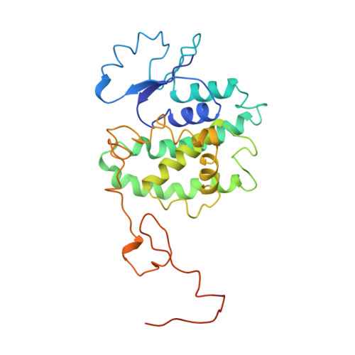
    A glutathione S-transferase fused with the nuclear matrix targeting signal (GST-NMTS) of AML-1/CBF-alpha2 has been crystallized by the vapor diffusion method using polyethylene glycol (PEG) as the precipitant. The NMTS is a 31-amino-acid signal peptide that can target the AML-1/CBF-alpha2 protein to the nuclear matrix. The crystal belongs to tetragonal space group P43212 with unit cell dimensions a = b = 93.4 A, c = 57.6 A. There is one GST-fusion protein per asymmetric unit. Crystals diffracted to at least 2.7 A and are appropriate for structure determination.


  • Organizational Affiliation
    • Program in Molecular Medicine, UMASS Medical Center, Worcester, Massachusetts, 01605, USA.

Macromolecule Content 

  • Total Structure Weight: 32.12 kDa 
  • Atom Count: 2,083 
  • Modeled Residue Count: 260 
  • Deposited Residue Count: 280 
  • Unique protein chains: 1

Macromolecules

Find similar proteins by:|  3D Structure
Entity ID: 1
MoleculeChains  Sequence LengthOrganismDetailsImage
PROTEIN (AML-1B)280Escherichia coliMutation(s): 0 
EC: 2.5.1.18
UniProt
Find proteins for P08515 (Schistosoma japonicum)
Explore P08515 
Go to UniProtKB:  P08515
Entity Groups
Sequence Clusters30% Identity50% Identity70% Identity90% Identity95% Identity100% Identity
UniProt GroupP08515
Sequence Annotations
Expand
Reference Sequence

Experimental Data & Validation

Experimental Data

  • Method: X-RAY DIFFRACTION
  • Resolution: 2.70 Å
  • R-Value Free:  0.310 (Depositor) 
  • R-Value Work:  0.209 (Depositor) 
Space Group: P 43 21 2
Unit Cell:
Length ( Å )Angle ( ˚ )
a = 93.4α = 90
b = 93.4β = 90
c = 57.6γ = 90
Software Package:
Software NamePurpose
AMoREphasing
X-PLORrefinement
DENZOdata reduction
SCALEPACKdata scaling

Structure Validation

View Full Validation Report


Entry History 

Deposition Data

Revision History  (Full details and data files)

  • Version 1.0: 1999-04-12
    Type: Initial release
  • Version 1.1: 2008-04-26
    Changes: Version format compliance
  • Version 1.2: 2011-07-13
    Changes: Derived calculations, Version format compliance
  • Version 1.3: 2023-08-09
    Changes: Data collection, Database references, Refinement description