1B8D

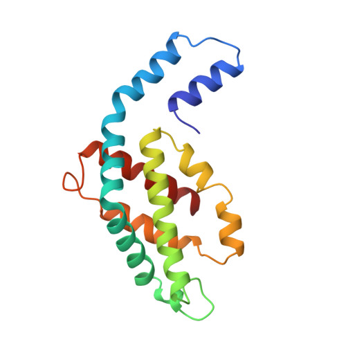
CRYSTAL STRUCTURE OF A PHYCOUROBILIN-CONTAINING PHYCOERYTHRIN


Experimental Data Snapshot

  • Method: X-RAY DIFFRACTION
  • Resolution: 1.90 Å
  • R-Value Free: 0.227 
  • R-Value Work: 0.175 

Starting Model: experimental
View more details

wwPDB Validation   3D Report Full Report


Ligand Structure Quality Assessment 

Created with Raphaël 2.3.0Worse 01 BetterLigand structure goodness of fit to experimental dataBest fitted PUBClick on this verticalbar to view detailsBest fitted PEBClick on this verticalbar to view details

This is version 1.3 of the entry. See complete history


Literature

Crystal structure of a phycourobilin-containing phycoerythrin at 1.90-A resolution.

Ritter, S.Hiller, R.G.Wrench, P.M.Welte, W.Diederichs, K.

(1999) J Struct Biol 126: 86-97

  • DOI: https://doi.org/10.1006/jsbi.1999.4106
  • Primary Citation of Related Structures:  
    1B8D

  • PubMed Abstract: 

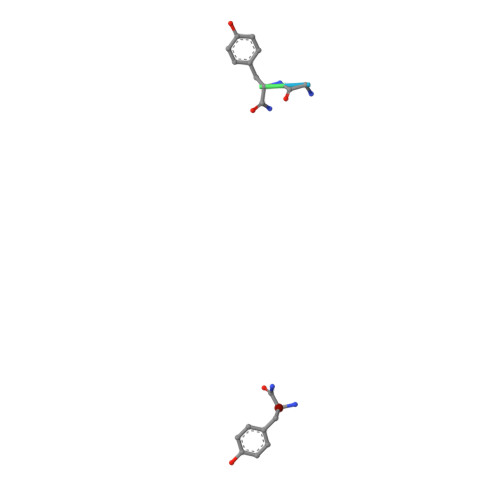
    The structure of R-phycoerythrin (R-PE) from the red alga Griffithsia monilis was solved at 1.90-A resolution by molecular replacement, using the atomic coordinates of cyanobacterial phycocyanin from Fremyella diplosiphon as a model. The crystallographic R factor for the final model is 17.5% (Rfree 22.7%) for reflections in the range 100-1.90 A. The model consists of an (alphabeta)2 dimer with an internal noncrystallographic dyad and a fragment of the gamma-polypeptide. The alpha-polypeptide (164 amino acid residues) has two covalently bound phycoerythrobilins at positions alpha82 and alpha139. The beta-polypeptide (177 residues) has two phycoerythrobilins bound to residues beta82 and beta158 and one phycourobilin covalently attached to rings A and D at residues beta50 and beta61, respectively. The electron density of the gamma-polypeptide is mostly averaged out by threefold crystallographic symmetry, but a dipeptide (Gly-Tyr) and one single Tyr could be modeled. These two tyrosine residues of the gamma-polypeptide are in close proximity to the phycoerythrobilins at position beta82 of two symmetry-related beta-polypeptides and are related by the same noncrystallographic dyad as the (alphabeta)2 dimer. Possible energy transfer pathways are discussed briefly.


  • Organizational Affiliation

    Institut für Biophysik und Strahlenbiologie, Universität Freiburg, Albertstrasse 23, Freiburg, D-79104, Germany.


Macromolecules
Find similar proteins by:  (by identity cutoff)  |  3D Structure
Entity ID: 1
MoleculeChains Sequence LengthOrganismDetailsImage
PROTEIN (RHODOPHYTAN PHYCOERYTHRIN (ALPHA CHAIN))A,
C [auth K]
164Griffithsia monilisMutation(s): 0 
UniProt
Find proteins for O36005 (Griffithsia monilis)
Explore O36005 
Go to UniProtKB:  O36005
Entity Groups  
Sequence Clusters30% Identity50% Identity70% Identity90% Identity95% Identity100% Identity
UniProt GroupO36005
Sequence Annotations
Expand
  • Reference Sequence
Find similar proteins by:  (by identity cutoff)  |  3D Structure
Entity ID: 2
MoleculeChains Sequence LengthOrganismDetailsImage
PROTEIN (RHODOPHYTAN PHYCOERYTHRIN (BETA CHAIN))B,
D [auth L]
177Griffithsia monilisMutation(s): 0 
UniProt
Find proteins for O36004 (Griffithsia monilis)
Explore O36004 
Go to UniProtKB:  O36004
Entity Groups  
Sequence Clusters30% Identity50% Identity70% Identity90% Identity95% Identity100% Identity
UniProt GroupO36004
Sequence Annotations
Expand
  • Reference Sequence

Find similar proteins by:  Sequence   |   3D Structure  

Entity ID: 3
MoleculeChains Sequence LengthOrganismDetailsImage
PROTEIN (RHODOPHYTAN PHYCOERYTHRIN (GAMMA CHAIN))E [auth G]6Griffithsia monilisMutation(s): 0 
Sequence Annotations
Expand
  • Reference Sequence
Small Molecules
Ligands 2 Unique
IDChains Name / Formula / InChI Key2D Diagram3D Interactions
PUB
Query on PUB

Download Ideal Coordinates CCD File 
J [auth B],
O [auth L]
PHYCOUROBILIN
C33 H42 N4 O6
KDCCOOGTVSRCHX-YYVBKQGDSA-N
PEB
Query on PEB

Download Ideal Coordinates CCD File 
F [auth A]
G [auth A]
H [auth B]
I [auth B]
K
PHYCOERYTHROBILIN
C33 H40 N4 O6
NKCBCVIFPXGHAV-WAVSMFBNSA-N
Modified Residues  1 Unique
IDChains TypeFormula2D DiagramParent
MEN
Query on MEN
B,
D [auth L]
L-PEPTIDE LINKINGC5 H10 N2 O3ASN
Experimental Data & Validation

Experimental Data

  • Method: X-RAY DIFFRACTION
  • Resolution: 1.90 Å
  • R-Value Free: 0.227 
  • R-Value Work: 0.175 
  • Space Group: H 3
Unit Cell:
Length ( Å )Angle ( ˚ )
a = 187.35α = 90
b = 187.35β = 90
c = 59.31γ = 120
Software Package:
Software NamePurpose
X-PLORmodel building
X-PLORrefinement
XDSdata reduction
XSCALEdata scaling
X-PLORphasing

Structure Validation

View Full Validation Report


Ligand Structure Quality Assessment 

Created with Raphaël 2.3.0Worse 01 BetterLigand structure goodness of fit to experimental dataBest fitted PUBClick on this verticalbar to view detailsBest fitted PEBClick on this verticalbar to view details

Entry History 

Deposition Data

Revision History  (Full details and data files)

  • Version 1.0: 1999-02-18
    Type: Initial release
  • Version 1.1: 2008-04-26
    Changes: Version format compliance
  • Version 1.2: 2011-07-13
    Changes: Version format compliance
  • Version 1.3: 2023-08-09
    Changes: Data collection, Database references, Derived calculations, Refinement description