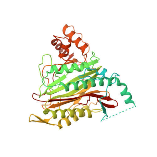
1B6A

HUMAN METHIONINE AMINOPEPTIDASE 2 COMPLEXED WITH TNP-470


Experimental Data Snapshot

  • Method: X-RAY DIFFRACTION
  • Resolution: 1.60 Å
  • R-Value Free: 0.216 
  • R-Value Work: 0.187 
  • R-Value Observed: 0.187 

wwPDB Validation   3D Report Full Report


Ligand Structure Quality Assessment 


This is version 1.5 of the entry. See complete history


Literature

Structure of human methionine aminopeptidase-2 complexed with fumagillin.

Liu, S.Widom, J.Kemp, C.W.Crews, C.M.Clardy, J.

(1998) Science 282: 1324-1327

  • DOI: https://doi.org/10.1126/science.282.5392.1324
  • Primary Citation of Related Structures:  
    1B59, 1B6A, 1BN5, 1BOA

  • PubMed Abstract: 

    The fungal metabolite fumagillin suppresses the formation of new blood vessels, and a fumagillin analog is currently in clinical trials as an anticancer agent. The molecular target of fumagillin is methionine aminopeptidase-2 (MetAP-2). A 1.8 A resolution crystal structure of free and inhibited human MetAP-2 shows a covalent bond formed between a reactive epoxide of fumagillin and histidine-231 in the active site of MetAP-2. Extensive hydrophobic and water-mediated polar interactions with other parts of fumagillin provide additional affinity. Fumagillin-based drugs inhibit MetAP-2 but not MetAP-1, and the three-dimensional structure also indicates the likely determinants of this specificity. The structural basis for fumagillin's potency and specificity forms the starting point for structure-based drug design.


  • Organizational Affiliation

    J. Clardy, Department of Chemistry and Chemical Biology, Baker Laboratory, Cornell University, Ithaca, NY 14853-1301, USA.


Macromolecules
Find similar proteins by:  (by identity cutoff)  |  3D Structure
Entity ID: 1
MoleculeChains Sequence LengthOrganismDetailsImage
METHIONINE AMINOPEPTIDASE478Homo sapiensMutation(s): 0 
EC: 3.4.11.18
UniProt & NIH Common Fund Data Resources
Find proteins for P50579 (Homo sapiens)
Explore P50579 
Go to UniProtKB:  P50579
PHAROS:  P50579
GTEx:  ENSG00000111142 
Entity Groups  
Sequence Clusters30% Identity50% Identity70% Identity90% Identity95% Identity100% Identity
UniProt GroupP50579
Sequence Annotations
Expand
  • Reference Sequence
Small Molecules
Ligands 2 Unique
IDChains Name / Formula / InChI Key2D Diagram3D Interactions
TN4
Query on TN4

Download Ideal Coordinates CCD File 
D [auth A](1R,2S,3S,4R)-4-hydroxy-2-methoxy-4-methyl-3-[(2R,3R)-2-methyl-3-(3-methylbut-2-en-1-yl)oxiran-2-yl]cyclohexyl (chloroacetyl)carbamate
C19 H30 Cl N O6
OUUSPVSSNHLIGE-AGYLCKTBSA-N
CO
Query on CO

Download Ideal Coordinates CCD File 
B [auth A],
C [auth A]
COBALT (II) ION
Co
XLJKHNWPARRRJB-UHFFFAOYSA-N
Experimental Data & Validation

Experimental Data

  • Method: X-RAY DIFFRACTION
  • Resolution: 1.60 Å
  • R-Value Free: 0.216 
  • R-Value Work: 0.187 
  • R-Value Observed: 0.187 
  • Space Group: C 2 2 21
Unit Cell:
Length ( Å )Angle ( ˚ )
a = 89.558α = 90
b = 99.06β = 90
c = 101.508γ = 90
Software Package:
Software NamePurpose
SYNCHROTRONdata collection
SCALAdata scaling
CNSrefinement
SOFTWAREdata reduction
CCP4data scaling
CNSphasing

Structure Validation

View Full Validation Report


Ligand Structure Quality Assessment 


Entry History 

Deposition Data

Revision History  (Full details and data files)

  • Version 1.0: 2000-01-13
    Type: Initial release
  • Version 1.1: 2008-03-24
    Changes: Version format compliance
  • Version 1.2: 2011-07-13
    Changes: Version format compliance
  • Version 1.3: 2011-08-31
    Changes: Non-polymer description
  • Version 1.4: 2019-11-20
    Changes: Advisory, Derived calculations
  • Version 1.5: 2023-08-02
    Changes: Database references, Derived calculations, Refinement description