1B57

CLASS II FRUCTOSE-1,6-BISPHOSPHATE ALDOLASE IN COMPLEX WITH PHOSPHOGLYCOLOHYDROXAMATE


Experimental Data Snapshot

  • Method: X-RAY DIFFRACTION
  • Resolution: 2.00 Å
  • R-Value Free: 0.230 
  • R-Value Work: 0.192 

wwPDB Validation   3D Report Full Report


Ligand Structure Quality Assessment 


This is version 1.3 of the entry. See complete history


Literature

The crystal structure of Escherichia coli class II fructose-1, 6-bisphosphate aldolase in complex with phosphoglycolohydroxamate reveals details of mechanism and specificity.

Hall, D.R.Leonard, G.A.Reed, C.D.Watt, C.I.Berry, A.Hunter, W.N.

(1999) J Mol Biol 287: 383-394

  • DOI: https://doi.org/10.1006/jmbi.1999.2609
  • Primary Citation of Related Structures:  
    1B57

  • PubMed Abstract: 

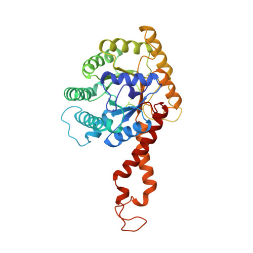
    The structure of a class II fructose-1,6-bisphosphate aldolase in complex with the substrate analogue and inhibitor phosphoglycolohydroxamate (PGH) has been determined using X-ray diffraction terms to a resolution of 2.0 A (1 A=0.1 nm). The crystals are trigonal, space group P3121 with a=b=78.24 A, c=289.69 A. The asymmetric unit is a homodimer of (alpha/beta)8 barrels and the model has refined to give R-work 19.2 %, R-free (based on 5 % of the data) 23.0 %. PGH resembles the ene-diolate transition state of the physiological substrate dihydroxyacetone phosphate. It is well ordered and bound in a deep polar cavity at the C-terminal end of the (alpha/beta)8 barrel, where it chelates the catalytic zinc ion using hydroxyl and enolate oxygen atoms. Trigonal bipyramidal coordination of the zinc ion is completed by three histidine residues. The complex network of hydrogen bonds at the catalytic centre is required to organise the position of key functional groups and metal ion ligands. A well-defined monovalent cation-binding site is observed following significant re-organisation of loop structures. This assists the formation of a phosphate-binding site on one side of the barrel that tethers PGH in the catalytic site. The positions of functional groups of substrate and putative interactions with key amino acid residues are identified. Knowledge of the complex structure complements the results of spectroscopic and site-directed mutagenesis studies, and contributes to our understanding of the mechanism and substrate specificity of this family of enzymes. A reaction mechanism distinct from that proposed for other class II aldolases is discussed. The results suggest that the class II aldolases should be sub-divided into two groups on the basis of both distinct folds and mechanism.


  • Organizational Affiliation

    Department of Biochemistry, University of Dundee, Dundee, DD1 5EH, UK.


Macromolecules
Find similar proteins by:  (by identity cutoff)  |  3D Structure
Entity ID: 1
MoleculeChains Sequence LengthOrganismDetailsImage
PROTEIN (FRUCTOSE-BISPHOSPHATE ALDOLASE II)
A, B
358Escherichia coliMutation(s): 0 
EC: 4.1.2.13
UniProt
Find proteins for P0AB71 (Escherichia coli (strain K12))
Explore P0AB71 
Go to UniProtKB:  P0AB71
Entity Groups  
Sequence Clusters30% Identity50% Identity70% Identity90% Identity95% Identity100% Identity
UniProt GroupP0AB71
Sequence Annotations
Expand
  • Reference Sequence
Small Molecules
Ligands 4 Unique
IDChains Name / Formula / InChI Key2D Diagram3D Interactions
PGH
Query on PGH

Download Ideal Coordinates CCD File 
I [auth A],
N [auth B]
PHOSPHOGLYCOLOHYDROXAMIC ACID
C2 H6 N O6 P
BAXHHWZKQZIJID-UHFFFAOYSA-N
ZN
Query on ZN

Download Ideal Coordinates CCD File 
C [auth A]
D [auth A]
E [auth A]
F [auth A]
J [auth B]
C [auth A],
D [auth A],
E [auth A],
F [auth A],
J [auth B],
K [auth B],
L [auth B]
ZINC ION
Zn
PTFCDOFLOPIGGS-UHFFFAOYSA-N
CL
Query on CL

Download Ideal Coordinates CCD File 
H [auth A]CHLORIDE ION
Cl
VEXZGXHMUGYJMC-UHFFFAOYSA-M
NA
Query on NA

Download Ideal Coordinates CCD File 
G [auth A],
M [auth B]
SODIUM ION
Na
FKNQFGJONOIPTF-UHFFFAOYSA-N
Binding Affinity Annotations 
IDSourceBinding Affinity
PGH PDBBind:  1B57 Ki: 10 (nM) from 1 assay(s)
Experimental Data & Validation

Experimental Data

  • Method: X-RAY DIFFRACTION
  • Resolution: 2.00 Å
  • R-Value Free: 0.230 
  • R-Value Work: 0.192 
  • Space Group: P 31 2 1
Unit Cell:
Length ( Å )Angle ( ˚ )
a = 78.237α = 90
b = 78.237β = 90
c = 289.687γ = 120
Software Package:
Software NamePurpose
AMoREphasing
REFMACrefinement
DENZOdata reduction
SCALEPACKdata scaling

Structure Validation

View Full Validation Report


Ligand Structure Quality Assessment 


Entry History 

Deposition Data

Revision History  (Full details and data files)

  • Version 1.0: 2000-01-07
    Type: Initial release
  • Version 1.1: 2008-04-27
    Changes: Version format compliance
  • Version 1.2: 2011-07-13
    Changes: Version format compliance
  • Version 1.3: 2023-08-09
    Changes: Data collection, Database references, Derived calculations, Refinement description