1B1G | pdb_00001b1g

SOLVATED REFINEMENT OF CA-LOADED CALBINDIN D9K


Experimental Data Snapshot

  • Method: SOLUTION NMR
  • Conformers Calculated: 10 
  • Conformers Submitted: 10 
  • Selection Criteria: ALL CONFORMERS USED 

wwPDB Validation 3D Report Full Report

Validation slider image for 1B1G

This is version 1.5 of the entry. See complete history

Literature

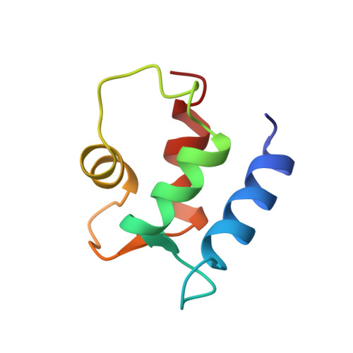
Protein solution structure calculations in solution: solvated molecular dynamics refinement of calbindin D9k.

Kordel, J.Pearlman, D.A.Chazin, W.J.

(1997) J Biomol NMR 10: 231-243

  • DOI: https://doi.org/10.1023/a:1018383102870
  • Primary Citation Related Structures: 
    1B1G

  • PubMed Abstract: 

    The three-dimensional solution structures of proteins determined with NMR-derived constraints are almost always calculated in vacuo. The solution structure of (Ca2+)2-calbindin D9k has been redetermined by new restrained molecular dynamics (MD) calculations that include Ca2+ ions and explicit solvent molecules. Four parallel sets of MD refinements were run to provide accurate comparisons of structures produced in vacuo, in vacuo with Ca2+ ions, and with two different protocols in a solvent bath with Ca2+ ions. The structural ensembles were analyzed in terms of structural definition, molecular energies, packing density, solvent-accessible surface, hydrogen bonds, and the coordination of calcium ions in the two binding loops. Refinement including Ca2+ ions and explicit solvent results in significant improvements in the precision and accuracy of the structure, particularly in the binding loops. These results are consistent with results previously obtained in free MD simulations of proteins in solution and show that the rMD refined NMR-derived solution structures of proteins, especially metalloproteins, can be significantly improved by these strategies.


  • Organizational Affiliation
    • Pharmacia & Upjohn, Stockholm, Sweden.

Macromolecule Content 

  • Total Structure Weight: 8.55 kDa 
  • Atom Count: 598 
  • Modeled Residue Count: 75 
  • Deposited Residue Count: 75 
  • Unique protein chains: 1

Macromolecules

Find similar proteins by:|  3D Structure
Entity ID: 1
MoleculeChains  Sequence LengthOrganismDetailsImage
PROTEIN (CALBINDIN D9K)75Bos taurusMutation(s): 0 
UniProt
Find proteins for P02633 (Bos taurus)
Explore P02633 
Go to UniProtKB:  P02633
Entity Groups
Sequence Clusters30% Identity50% Identity70% Identity90% Identity95% Identity100% Identity
UniProt GroupP02633
Sequence Annotations
Expand
Reference Sequence

Small Molecules

Experimental Data & Validation

Experimental Data

  • Method: SOLUTION NMR
  • Conformers Calculated: 10 
  • Conformers Submitted: 10 
  • Selection Criteria: ALL CONFORMERS USED 

Structure Validation

View Full Validation Report


Entry History 

Deposition Data

Revision History  (Full details and data files)

  • Version 1.0: 1998-11-25
    Type: Initial release
  • Version 1.1: 2008-04-26
    Changes: Version format compliance
  • Version 1.2: 2011-07-13
    Changes: Version format compliance
  • Version 1.3: 2011-08-10
    Changes: Other
  • Version 1.4: 2023-12-27
    Changes: Data collection, Database references, Derived calculations
  • Version 1.5: 2025-03-12
    Changes: Database references, Structure summary