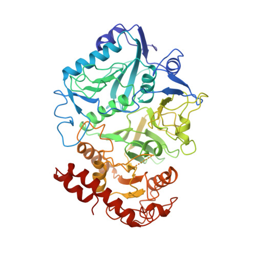
1AYL

PHOSPHOENOLPYRUVATE CARBOXYKINASE


Experimental Data Snapshot

  • Method: X-RAY DIFFRACTION
  • Resolution: 1.80 Å
  • R-Value Free: 0.234 
  • R-Value Work: 0.195 
  • R-Value Observed: 0.195 

wwPDB Validation   3D Report Full Report


This is version 1.5 of the entry. See complete history


Literature

Snapshot of an enzyme reaction intermediate in the structure of the ATP-Mg2+-oxalate ternary complex of Escherichia coli PEP carboxykinase.

Tari, L.W.Matte, A.Pugazhenthi, U.Goldie, H.Delbaere, L.T.

(1996) Nat Struct Biol 3: 355-363

  • DOI: https://doi.org/10.1038/nsb0496-355
  • Primary Citation of Related Structures:  
    1AYL

  • PubMed Abstract: 

    We report the 1.8 A crystal structure of adenosine triphosphate (ATP)-magnesium-oxalate bound phosphoenolpyruvate carboxykinase (PCK) from Escherichia coli. ATP binding induces a 20 degree hinge-like rotation of the N- and C-terminal domains which closes the active-site cleft. PCK possesses a novel nucleotide-binding fold, particularly in the adenine-binding region, where the formation of a cis backbone torsion angle in a loop glycine residue promotes intimate contacts between the adenine-binding loop and adenine, while stabilizing a syn conformation of the base. This complex represents a reaction intermediate analogue along the pathway of the conversion of oxaloacetate to phosphoenolpyruvate, and provides insight into the mechanistic details of the chemical reaction catalysed by this enzyme.


  • Organizational Affiliation

    Department of Biochemistry, University of Saskatchewan, Saskatoon, Saskatchewan, Canada.


Macromolecules
Find similar proteins by:  (by identity cutoff)  |  3D Structure
Entity ID: 1
MoleculeChains Sequence LengthOrganismDetailsImage
PHOSPHOENOLPYRUVATE CARBOXYKINASE541Escherichia coli K-12Mutation(s): 0 
EC: 4.1.1.49
UniProt
Find proteins for P22259 (Escherichia coli (strain K12))
Explore P22259 
Go to UniProtKB:  P22259
Entity Groups  
Sequence Clusters30% Identity50% Identity70% Identity90% Identity95% Identity100% Identity
UniProt GroupP22259
Sequence Annotations
Expand
  • Reference Sequence
Small Molecules
Ligands 3 Unique
IDChains Name / Formula / InChI Key2D Diagram3D Interactions
ATP
Query on ATP

Download Ideal Coordinates CCD File 
D [auth A]ADENOSINE-5'-TRIPHOSPHATE
C10 H16 N5 O13 P3
ZKHQWZAMYRWXGA-KQYNXXCUSA-N
OXL
Query on OXL

Download Ideal Coordinates CCD File 
B [auth A]OXALATE ION
C2 O4
MUBZPKHOEPUJKR-UHFFFAOYSA-L
MG
Query on MG

Download Ideal Coordinates CCD File 
C [auth A]MAGNESIUM ION
Mg
JLVVSXFLKOJNIY-UHFFFAOYSA-N
Experimental Data & Validation

Experimental Data

  • Method: X-RAY DIFFRACTION
  • Resolution: 1.80 Å
  • R-Value Free: 0.234 
  • R-Value Work: 0.195 
  • R-Value Observed: 0.195 
  • Space Group: C 1 2 1
Unit Cell:
Length ( Å )Angle ( ˚ )
a = 127.51α = 90
b = 96.69β = 96.63
c = 46.96γ = 90
Software Package:
Software NamePurpose
X-PLORrefinement
WEISdata reduction

Structure Validation

View Full Validation Report


Entry History 

Deposition Data

Revision History  (Full details and data files)

  • Version 1.0: 1997-01-11
    Type: Initial release
  • Version 1.1: 2008-03-24
    Changes: Version format compliance
  • Version 1.2: 2011-07-13
    Changes: Source and taxonomy, Version format compliance
  • Version 1.3: 2017-11-29
    Changes: Derived calculations, Other
  • Version 1.4: 2018-03-21
    Changes: Data collection
  • Version 1.5: 2024-02-07
    Changes: Data collection, Database references, Derived calculations