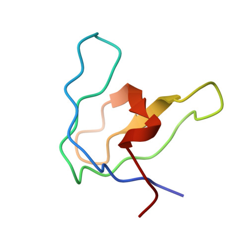
The crystal structure of HIV-1 Nef protein bound to the Fyn kinase SH3 domain suggests a role for this complex in altered T cell receptor signaling.
Arold, S., Franken, P., Strub, M.P., Hoh, F., Benichou, S., Benarous, R., Dumas, C.(1997) Structure 5: 1361-1372
- PubMed: 9351809 Search on PubMed
- DOI: https://doi.org/10.1016/s0969-2126(97)00286-4
- Primary Citation Related Structures:
1AVV, 1AVZ - PubMed Abstract:
Human immunodeficiency virus (HIV) Nef protein accelerates virulent progression of acquired immunodeficiency syndrome (AIDS) by its interaction with specific cellular proteins involved in signal transduction and host cell activation. Nef has been shown to bind specifically to a subset of the Src family of kinases. The structures of free Nef and Nef bound to Src homology region 3 (SH3) domain are important for the elucidation of how the affinity and specificity for the Src kinase family SH3 domains are achieved, and also for the development of potential drugs and vaccines against AIDS. We have determined the crystal structures of the conserved core of HIV-1 Nef protein alone and in complex with the wild-type SH3 domain of the p59fyn protein tyrosine kinase (Fyn), at 3.0 A resolution. Comparison of the bound and unbound Nef structures revealed that a proline-rich motif (Pro-x-x-Pro), which is implicated in SH3 binding, is partially disordered in the absence of the binding partner; this motif only fully adopts a left-handed polyproline type II helix conformation upon complex formation with the Fyn SH3 domain. In addition, the structures show how an arginine residue (Arg77) of Nef interacts with Asp 100 of the so-called RT loop within the Fyn SH3 domain, and triggers a hydrogen-bond rearrangement which allows the loop to adapt to complement the Nef surface. The Arg96 residue of the Fyn SH3 domain is specifically accommodated in the same hydrophobic pocket of Nef as the isoleucine residue of a previously described Fyn SH3 (Arg96-->lle) mutant that binds to Nef with higher affinity than the wild type. The three-dimensional structures support evidence that the Nef-Fyn complex forms in vivo and may have a crucial role in the T cell perturbating action of Nef by altering T cell receptor signaling. The structures of bound and unbound Nef reveal that the multivalency of SH3 binding may be achieved by a ligand induced flexibility in the RT loop. The structures suggest possible targets for the design of inhibitors which specifically block Nef-SH3 interactions.
- Centre de Biochimie Structurale, UMR C9955 CNRS, U414 INSERM, Université Montpellier I, Faculté de Pharmacie, France.
Organizational Affiliation: