The solution structure of the complex of Lactobacillus casei dihydrofolate reductase with methotrexate.
Gargaro, A.R., Soteriou, A., Frenkiel, T.A., Bauer, C.J., Birdsall, B., Polshakov, V.I., Barsukov, I.L., Roberts, G.C., Feeney, J.(1998) J Mol Biology 277: 119-134
- PubMed: 9514736 Search on PubMed
- DOI: https://doi.org/10.1006/jmbi.1997.1560
- Primary Citation Related Structures:
1AO8 - PubMed Abstract:
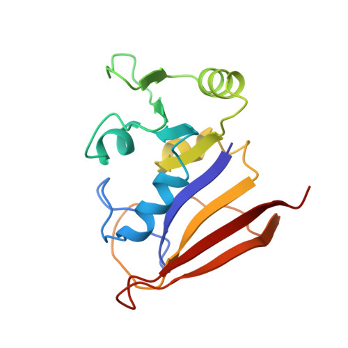
We have determined the three-dimensional solution structure of the complex of Lactobacillus casei dihydrofolate reductase (18.3 kDa, 162 amino acid residues) formed with the anticancer drug methotrexate using 2531 distance, 361 dihedral angle and 48 hydrogen bond restraints obtained from analysis of multidimensional NMR spectra. Simulated annealing calculations produced a family of 21 structures fully consistent with the constraints. The structure has four alpha-helices and eight beta-strands with two other regions, comprising residues 11 to 14 and 126 to 127, also interacting with each other in a beta-sheet manner. The methotrexate binding site is very well defined and the structure around its glutamate moiety was improved by including restraints reflecting the previously determined specific interactions between the glutamate alpha-carboxylate group with Arg57 and the gamma-carboxylate group with His28. The overall fold of the binary complex in solution is very similar to that observed in the X-ray studies of the ternary complex of L. casei dihydrofolate reductase formed with methotrexate and NADPH (the structures of the binary and ternary complexes have a root-mean-square difference over the backbone atoms of 0.97 A). Thus no major conformational change takes place when NADPH binds to the binary complex. In the binary complex, the loop comprising residues 9 to 23 which forms part of the active site has been shown to be in the "closed" conformation as defined by M. R. Sawaya & J. Kraut, who considered the corresponding loops in crystal structures of complexes of dihydrofolate reductases from several organisms. Thus the absence of the NADPH does not result in the "occluded" form of the loop as seen in crystal studies of some other dihydrofolate reductases in the absence of coenzyme. Some regions of the structure in the binary complex which form interaction sites for NADPH are less well defined than other regions. However, in general terms, the NADPH binding site appears to be essentially pre-formed in the binary complex. This may contribute to the tighter binding of coenzyme in the presence of methotrexate.
- Division of Molecular Structure, National Institute for Medical Research, London, UK.
Organizational Affiliation: