1ANV

ADENOVIRUS 5 DBP/URANYL FLUORIDE SOAK


Experimental Data Snapshot

  • Method: X-RAY DIFFRACTION
  • Resolution: 2.70 Å
  • R-Value Free: 0.310 
  • R-Value Work: 0.204 

wwPDB Validation   3D Report Full Report


This is version 1.4 of the entry. See complete history


Literature

Conformational change of the adenovirus DNA-binding protein induced by soaking crystals with K3UO2F5 solutions.

Kanellopoulos, P.N.Tsernoglou, D.van der Vliet, P.C.Tucker, P.A.

(1996) Acta Crystallogr D Biol Crystallogr 52: 942-945

  • DOI: https://doi.org/10.1107/S0907444996005525
  • Primary Citation of Related Structures:  
    1ANV

  • PubMed Abstract: 

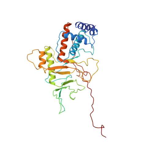
    Soaking crystals of the C-terminal DNA-binding domain of the adenovirus single-stranded DNA-binding protein with a buffer containing K(3)UO(2)F(5) results in a 9% change of the crystallographic c axis without destruction of the crystals or appreciable loss of resolution. The crystals belong to space group P2(1)2(1)2(1) with a = 79.7, b = 75.6 and c = 60.6 A. The three-dimensional structure has been refined to 2.7 A with a crystallographic R factor of 0.206. Antiparallel chains of protein molecules running through the entire crystal are linked by uranyl ions. The relative orientation of protein monomers is flexible, even in the crystalline state, and allows changes in the packing of the protein chains.


  • Organizational Affiliation

    Structural Biology Programme, European Molecular Biology Laboratory, Heidelberg, Germany.


Macromolecules
Find similar proteins by:  (by identity cutoff)  |  3D Structure
Entity ID: 1
MoleculeChains Sequence LengthOrganismDetailsImage
ADENOVIRUS SINGLE-STRANDED DNA-BINDING PROTEIN356Human adenovirus 5Mutation(s): 0 
UniProt
Find proteins for P03265 (Human adenovirus C serotype 5)
Explore P03265 
Go to UniProtKB:  P03265
Entity Groups  
Sequence Clusters30% Identity50% Identity70% Identity90% Identity95% Identity100% Identity
UniProt GroupP03265
Sequence Annotations
Expand
  • Reference Sequence
Experimental Data & Validation

Experimental Data

  • Method: X-RAY DIFFRACTION
  • Resolution: 2.70 Å
  • R-Value Free: 0.310 
  • R-Value Work: 0.204 
  • Space Group: P 21 21 21
Unit Cell:
Length ( Å )Angle ( ˚ )
a = 79.7α = 90
b = 75.6β = 90
c = 60.6γ = 90
Software Package:
Software NamePurpose
AMoREphasing
TNTrefinement
XDSdata reduction
XDSdata scaling

Structure Validation

View Full Validation Report


Entry History 

Deposition Data

Revision History  (Full details and data files)

  • Version 1.0: 1997-02-12
    Type: Initial release
  • Version 1.1: 2008-03-24
    Changes: Version format compliance
  • Version 1.2: 2011-07-13
    Changes: Version format compliance
  • Version 1.3: 2018-04-04
    Changes: Data collection, Structure summary
  • Version 1.4: 2023-08-02
    Changes: Database references, Derived calculations, Refinement description