1AM9

HUMAN SREBP-1A BOUND TO LDL RECEPTOR PROMOTER


Experimental Data Snapshot

  • Method: X-RAY DIFFRACTION
  • Resolution: 2.30 Å
  • R-Value Free: 0.278 
  • R-Value Work: 0.219 
  • R-Value Observed: 0.219 

wwPDB Validation   3D Report Full Report


This is version 1.4 of the entry. See complete history


Literature

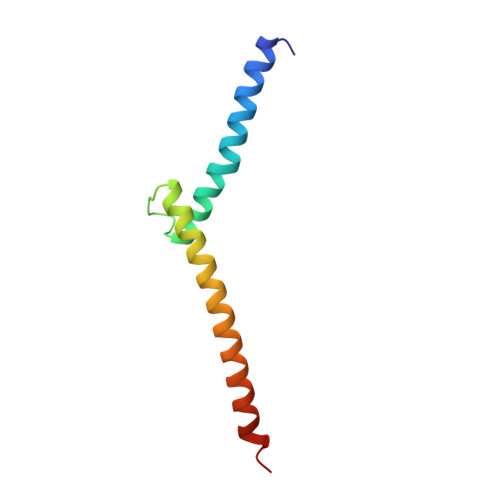
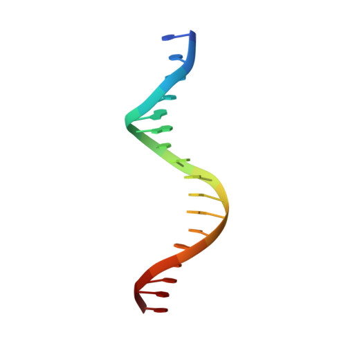
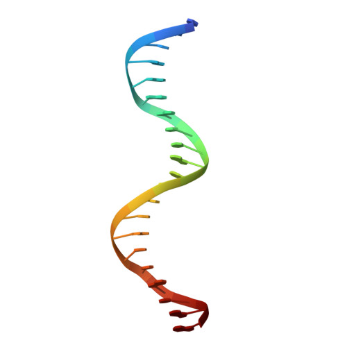
Co-crystal structure of sterol regulatory element binding protein 1a at 2.3 A resolution.

Parraga, A.Bellsolell, L.Ferre-D'Amare, A.R.Burley, S.K.

(1998) Structure 6: 661-672

  • DOI: https://doi.org/10.1016/s0969-2126(98)00067-7
  • Primary Citation of Related Structures:  
    1AM9

  • PubMed Abstract: 

    The sterol regulatory element binding proteins (SREBPs) are helix-loop-helix transcriptional activators that control expression of genes encoding proteins essential for cholesterol biosynthesis/uptake and fatty acid biosynthesis. Unlike helix-loop-helix proteins that recognize symmetric E-boxes (5'-CANNTG-3'), the SREBPs have a tyrosine instead of a conserved arginine in their basic regions. This difference allows recognition of an asymmetric sterol regulatory element (StRE, 5'-ATCACCCAC-3').


  • Organizational Affiliation

    Laboratories of Molecular Biophysics, Rockefeller University, New York, NY 10021, USA.


Macromolecules

Find similar proteins by:  (by identity cutoff)  |  3D Structure
Entity ID: 3
MoleculeChains Sequence LengthOrganismDetailsImage
PROTEIN (STEROL REGULATORY ELEMENT BINDING PROTEIN 1A)E [auth A],
F [auth B],
G [auth C],
H [auth D]
82Homo sapiensMutation(s): 0 
UniProt & NIH Common Fund Data Resources
Find proteins for P36956 (Homo sapiens)
Explore P36956 
Go to UniProtKB:  P36956
PHAROS:  P36956
GTEx:  ENSG00000072310 
Entity Groups  
Sequence Clusters30% Identity50% Identity70% Identity90% Identity95% Identity100% Identity
UniProt GroupP36956
Sequence Annotations
Expand
  • Reference Sequence

Find similar nucleic acids by:  Sequence   |   3D Structure  

Entity ID: 1
MoleculeChains LengthOrganismImage
DNA (5'-D(*TP*TP*GP*CP*AP*GP*TP*GP*GP*GP*GP*TP*GP*AP*TP*CP*T )-3')A [auth E],
C [auth G]
17N/A
Sequence Annotations
Expand
  • Reference Sequence

Find similar nucleic acids by:  Sequence   |   3D Structure  

Entity ID: 2
MoleculeChains LengthOrganismImage
DNA (5'-D(*CP*AP*TP*GP*AP*GP*AP*TP*CP*AP*CP*CP*CP*CP*AP*CP*T P*GP*CP*AP*A)-3')B [auth F],
D [auth H]
21N/A
Sequence Annotations
Expand
  • Reference Sequence
Small Molecules
Ligands 1 Unique
IDChains Name / Formula / InChI Key2D Diagram3D Interactions
MG
Query on MG

Download Ideal Coordinates CCD File 
I [auth B],
J [auth C]
MAGNESIUM ION
Mg
JLVVSXFLKOJNIY-UHFFFAOYSA-N
Experimental Data & Validation

Experimental Data

  • Method: X-RAY DIFFRACTION
  • Resolution: 2.30 Å
  • R-Value Free: 0.278 
  • R-Value Work: 0.219 
  • R-Value Observed: 0.219 
  • Space Group: P 61 2 2
Unit Cell:
Length ( Å )Angle ( ˚ )
a = 94.63α = 90
b = 94.63β = 90
c = 459.1γ = 120
Software Package:
Software NamePurpose
AMoREphasing
X-PLORrefinement
DENZOdata reduction
SCALEPACKdata scaling

Structure Validation

View Full Validation Report


Entry History 

Deposition Data

Revision History  (Full details and data files)

  • Version 1.0: 1998-07-10
    Type: Initial release
  • Version 1.1: 2008-05-22
    Changes: Version format compliance
  • Version 1.2: 2011-07-13
    Changes: Version format compliance
  • Version 1.3: 2021-02-03
    Changes: Database references, Derived calculations, Structure summary
  • Version 1.4: 2024-02-07
    Changes: Data collection, Database references