1AJN

PENICILLIN ACYLASE COMPLEXED WITH P-NITROPHENYLACETIC ACID


Experimental Data Snapshot

  • Method: X-RAY DIFFRACTION
  • Resolution: 2.36 Å
  • R-Value Free: 0.218 
  • R-Value Work: 0.152 

wwPDB Validation   3D Report Full Report


Ligand Structure Quality Assessment 


This is version 1.3 of the entry. See complete history


Literature

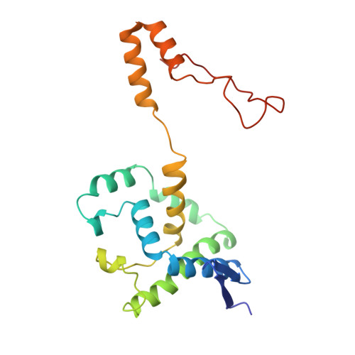
Ligand-induced conformational change in penicillin acylase.

Done, S.H.Brannigan, J.A.Moody, P.C.E.Hubbard, R.E.

(1998) J Mol Biol 284: 463-475

  • DOI: https://doi.org/10.1006/jmbi.1998.2180
  • Primary Citation of Related Structures:  
    1AI4, 1AI5, 1AI6, 1AI7, 1AJN, 1AJP, 1AJQ

  • PubMed Abstract: 

    The enzyme penicillin acylase (penicillin amidohydrolase EC 3.5.1. 11) catalyses the cleavage of the amide bond in the benzylpenicillin (penicillin G) side-chain to produce phenylacetic acid and 6-aminopenicillanic acid (6-APA). The enzyme is of great pharmaceutical importance, as the product 6-APA is the starting point for the synthesis of many semi-synthetic penicillin antibiotics. Studies have shown that the enzyme is specific for hydrolysis of phenylacetamide derivatives, but is more tolerant of features in the rest of the substrate. It is this property that has led to many other applications for the enzyme, and greater knowledge of the enzyme's structure and specificity could facilitate engineering of the enzyme, enhancing its potential for chemical and industrial applications. An extensive study of the binding of a series of phenylacetic acid derivatives has been carried out. A measure of the relative degree of inhibition of the enzyme by each of the compounds has been obtained using a competitive inhibition assay, and the structures of a number of these complexes have been determined by X-ray crystallography. The structures reveal a clear rationale for the observed kinetic results, but show also that some of the ligands cause a conformational change within the binding pocket. This change can generally be understood in terms of the size and orientation of the ligand within the active site.The results reveal that ligand binding in penicillin acylase is facilitated by certain amino acid residues that can adopt two distinct, energetically favourable positions in order to accommodate a variety of compounds within the active site. The structures of these complexes provide evidence for conformational changes in the substrate-binding region that may act as a switch in the mechanism of autocatalytic processing of this enzyme.


  • Organizational Affiliation

    Department of Chemistry, University of York, Heslington, YO1 5DD, UK. done@yorvic.york.ac.uk


Macromolecules
Find similar proteins by:  (by identity cutoff)  |  3D Structure
Entity ID: 1
MoleculeChains Sequence LengthOrganismDetailsImage
PENICILLIN AMIDOHYDROLASE209Escherichia coliMutation(s): 0 
EC: 3.5.1.11
UniProt
Find proteins for P06875 (Escherichia coli)
Explore P06875 
Go to UniProtKB:  P06875
Entity Groups  
Sequence Clusters30% Identity50% Identity70% Identity90% Identity95% Identity100% Identity
UniProt GroupP06875
Sequence Annotations
Expand
  • Reference Sequence
Find similar proteins by:  (by identity cutoff)  |  3D Structure
Entity ID: 2
MoleculeChains Sequence LengthOrganismDetailsImage
PENICILLIN AMIDOHYDROLASE557Escherichia coliMutation(s): 0 
EC: 3.5.1.11
UniProt
Find proteins for P06875 (Escherichia coli)
Explore P06875 
Go to UniProtKB:  P06875
Entity Groups  
Sequence Clusters30% Identity50% Identity70% Identity90% Identity95% Identity100% Identity
UniProt GroupP06875
Sequence Annotations
Expand
  • Reference Sequence
Small Molecules
Ligands 2 Unique
IDChains Name / Formula / InChI Key2D Diagram3D Interactions
AAN
Query on AAN

Download Ideal Coordinates CCD File 
D [auth B]2-(4-NITROPHENYL)ACETIC ACID
C8 H7 N O4
YBADLXQNJCMBKR-UHFFFAOYSA-N
CA
Query on CA

Download Ideal Coordinates CCD File 
C [auth B]CALCIUM ION
Ca
BHPQYMZQTOCNFJ-UHFFFAOYSA-N
Binding Affinity Annotations 
IDSourceBinding Affinity
AAN PDBBind:  1AJN Ki: 2.32e+6 (nM) from 1 assay(s)
Binding MOAD:  1AJN Ki: 2.32e+6 (nM) from 1 assay(s)
Experimental Data & Validation

Experimental Data

  • Method: X-RAY DIFFRACTION
  • Resolution: 2.36 Å
  • R-Value Free: 0.218 
  • R-Value Work: 0.152 
  • Space Group: P 1
Unit Cell:
Length ( Å )Angle ( ˚ )
a = 52.12α = 100.2
b = 65.08β = 111.44
c = 76.3γ = 105.81
Software Package:
Software NamePurpose
REFMACrefinement
DENZOdata reduction
CCP4data scaling
ROTAVATAdata scaling

Structure Validation

View Full Validation Report


Ligand Structure Quality Assessment 


Entry History 

Deposition Data

  • Released Date: 1997-11-12 
  • Deposition Author(s): Done, S.H.

Revision History  (Full details and data files)

  • Version 1.0: 1997-11-12
    Type: Initial release
  • Version 1.1: 2008-03-03
    Changes: Version format compliance
  • Version 1.2: 2011-07-13
    Changes: Version format compliance
  • Version 1.3: 2024-02-07
    Changes: Data collection, Database references, Derived calculations, Other, Refinement description