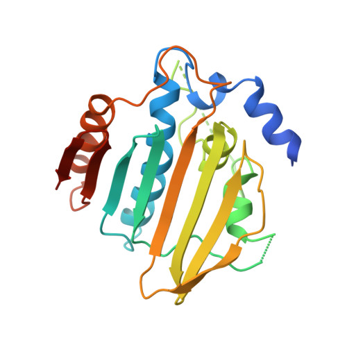
1AJ6

NOVOBIOCIN-RESISTANT MUTANT (R136H) OF THE N-TERMINAL 24 KDA FRAGMENT OF DNA GYRASE B COMPLEXED WITH NOVOBIOCIN AT 2.3 ANGSTROMS RESOLUTION


Experimental Data Snapshot

  • Method: X-RAY DIFFRACTION
  • Resolution: 2.30 Å
  • R-Value Free: 0.290 
  • R-Value Work: 0.195 
  • R-Value Observed: 0.206 

Starting Model: experimental
View more details

wwPDB Validation   3D Report Full Report


Ligand Structure Quality Assessment 


This is version 1.5 of the entry. See complete history


Literature

The entropic penalty of ordered water accounts for weaker binding of the antibiotic novobiocin to a resistant mutant of DNA gyrase: a thermodynamic and crystallographic study.

Holdgate, G.A.Tunnicliffe, A.Ward, W.H.Weston, S.A.Rosenbrock, G.Barth, P.T.Taylor, I.W.Pauptit, R.A.Timms, D.

(1997) Biochemistry 36: 9663-9673

  • DOI: https://doi.org/10.1021/bi970294+
  • Primary Citation of Related Structures:  
    1AJ6

  • PubMed Abstract: 

    Novobiocin is an antibiotic which binds to a 24 kDa fragment from the B subunit of DNA gyrase. Naturally occurring resistance arises from mutation of Arg-136 which hydrogen bonds to the coumarin ring of novobiocin. We have applied calorimetry to characterize the binding of novobiocin to wild-type and R136H mutant 24 kDa fragments. Upon mutation, the Kd increases from 32 to 1200 nM at 300 K. The enthalpy of binding is more favorable for the mutant (DeltaH degrees shifts from -12.1 to -17.5 kcal/mol), and the entropy of binding is much less favorable (TDeltaS degrees changes from -1.8 to -9.4 kcal/mol). Both of these changes are in the direction opposite to that expected if the loss of the Arg residue reduces hydrogen bonding. The change in heat capacity at constant pressure upon binding (DeltaCp) shifts from -295 to -454 cal mol-1 K-1. We also report the crystal structure, at 2.3 A resolution, of a complex between the R136H 24 kDa fragment and novobiocin. Although the change in DeltaCp often would be interpreted as reflecting increased burial of hydrophobic surface on binding, this structure reveals a small decrease. Furthermore, an ordered water molecule is sequestered into the volume vacated by removal of the guanidinium group. There are large discrepancies when the measured thermodynamic parameters are compared to those estimated from the structural data using empirical relationships. These differences seem to arise from the effects of sequestering ordered water molecules upon complexation. The water-mediated hydrogen bonds linking novobiocin to the mutant protein make a favorable enthalpic contribution, whereas the immobilization of the water leads to an entropic cost and a reduction in the heat capacity of the system. Such a negative contribution to DeltaCp, DeltaH degrees , and TDeltaS degrees appears to be a general property of water molecules that are sequestered when ligands bind to proteins.


  • Organizational Affiliation

    ZENECA Pharmaceuticals, Macclesfield, Cheshire, U.K.


Macromolecules
Find similar proteins by:  (by identity cutoff)  |  3D Structure
Entity ID: 1
MoleculeChains Sequence LengthOrganismDetailsImage
GYRASE219Escherichia coliMutation(s): 1 
Gene Names: GYRB
EC: 5.99.1.3 (PDB Primary Data), 5.6.2.2 (UniProt)
UniProt
Find proteins for P0AES6 (Escherichia coli (strain K12))
Explore P0AES6 
Go to UniProtKB:  P0AES6
Entity Groups  
Sequence Clusters30% Identity50% Identity70% Identity90% Identity95% Identity100% Identity
UniProt GroupP0AES6
Sequence Annotations
Expand
  • Reference Sequence
Small Molecules
Ligands 1 Unique
IDChains Name / Formula / InChI Key2D Diagram3D Interactions
NOV
Query on NOV

Download Ideal Coordinates CCD File 
B [auth A]NOVOBIOCIN
C31 H36 N2 O11
YJQPYGGHQPGBLI-KGSXXDOSSA-N
Binding Affinity Annotations 
IDSourceBinding Affinity
NOV BindingDB:  1AJ6 Ki: min: 7, max: 14 (nM) from 2 assay(s)
Kd: 7 (nM) from 1 assay(s)
IC50: min: 24, max: 408 (nM) from 5 assay(s)
-TΔS: 9.09 (kJ/mol) from 1 assay(s)
ΔH: -5.18e+1 (kJ/mol) from 1 assay(s)
ΔG: -4.27e+1 (kJ/mol) from 1 assay(s)
PDBBind:  1AJ6 Kd: 1200 (nM) from 1 assay(s)
Experimental Data & Validation

Experimental Data

  • Method: X-RAY DIFFRACTION
  • Resolution: 2.30 Å
  • R-Value Free: 0.290 
  • R-Value Work: 0.195 
  • R-Value Observed: 0.206 
  • Space Group: P 21 21 21
Unit Cell:
Length ( Å )Angle ( ˚ )
a = 39.5α = 90
b = 47.72β = 90
c = 114.41γ = 90
Software Package:
Software NamePurpose
CCP4model building
TNTrefinement
MOSFLMdata reduction
CCP4data scaling
CCP4phasing

Structure Validation

View Full Validation Report


Ligand Structure Quality Assessment 


Entry History 

Deposition Data

Revision History  (Full details and data files)

  • Version 1.0: 1998-05-20
    Type: Initial release
  • Version 1.1: 2008-03-03
    Changes: Version format compliance
  • Version 1.2: 2011-07-13
    Changes: Non-polymer description, Version format compliance
  • Version 1.3: 2021-11-03
    Changes: Database references, Derived calculations, Other
  • Version 1.4: 2024-02-07
    Changes: Data collection
  • Version 1.5: 2024-04-03
    Changes: Refinement description