1AIH | pdb_00001aih

CATALYTIC DOMAIN OF BACTERIOPHAGE HP1 INTEGRASE


Experimental Data Snapshot

  • Method: X-RAY DIFFRACTION
  • Resolution: 2.50 Å
  • R-Value Free: 
    0.272 (Depositor) 
  • R-Value Work: 
    0.209 (Depositor) 
  • R-Value Observed: 
    0.209 (Depositor) 

wwPDB Validation 3D Report Full Report

Validation slider image for 1AIH

This is version 1.3 of the entry. See complete history

Literature

Molecular organization in site-specific recombination: the catalytic domain of bacteriophage HP1 integrase at 2.7 A resolution.

Hickman, A.B.Waninger, S.Scocca, J.J.Dyda, F.

(1997) Cell 89: 227-237

  • DOI: https://doi.org/10.1016/s0092-8674(00)80202-0
  • Primary Citation Related Structures: 
    1AIH

  • PubMed Abstract: 

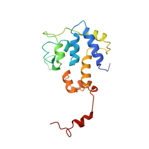
    HP1 integrase promotes site-specific recombination of the HP1 genome into that of Haemophilus influenzae. The isolated C-terminal domain (residues 165-337) of the protein interacts with the recombination site and contains the four catalytic residues conserved in the integrase family. This domain represents a novel fold consisting principally of well-packed alpha helices, a surface beta sheet, and an ordered 17-residue C-terminal tail. The conserved triad of basic residues and the active-site tyrosine are contributed by a single monomer and occupy fixed positions in a defined active-site cleft. Dimers are formed by mutual interactions of the tail of one monomer with an adjacent monomer; this orients active-site clefts antiparallel to each other.


  • Organizational Affiliation
    • Laboratory of Molecular Biology, National Institute of Diabetes and Digestive and Kidney Diseases, National Institutes of Health, Bethesda, Maryland 20892, USA.

Macromolecule Content 

  • Total Structure Weight: 78.72 kDa 
  • Atom Count: 5,545 
  • Modeled Residue Count: 676 
  • Deposited Residue Count: 680 
  • Unique protein chains: 1

Macromolecules

Find similar proteins by:|  3D Structure
Entity ID: 1
MoleculeChains  Sequence LengthOrganismDetailsImage
HP1 INTEGRASE
A, B, C, D
170Hpunavirus HP1Mutation(s): 0 
Gene Names: GENBANK U24159
EC: 3.1 (UniProt), 2.7.7 (UniProt)
UniProt
Find proteins for P21442 (Haemophilus phage HP1 (strain HP1c1))
Explore P21442 
Go to UniProtKB:  P21442
Entity Groups
Sequence Clusters30% Identity50% Identity70% Identity90% Identity95% Identity100% Identity
UniProt GroupP21442
Sequence Annotations
Expand
Reference Sequence

Small Molecules

Ligands 2 Unique
IDChains Name / Formula / InChI Key2D Diagram3D Interactions
SO4

Query on SO4


Download:Ideal Coordinates CCD File
E [auth A]
F [auth A]
G [auth A]
I [auth B]
J [auth B]
E [auth A],
F [auth A],
G [auth A],
I [auth B],
J [auth B],
L [auth C],
O [auth D],
P [auth D]
SULFATE ION
O4 S
QAOWNCQODCNURD-UHFFFAOYSA-L
MG

Query on MG


Download:Ideal Coordinates CCD File
H [auth A],
K [auth B],
M [auth C],
N [auth C],
Q [auth D]
MAGNESIUM ION
Mg
JLVVSXFLKOJNIY-UHFFFAOYSA-N

Experimental Data & Validation

Experimental Data

  • Method: X-RAY DIFFRACTION
  • Resolution: 2.50 Å
  • R-Value Free:  0.272 (Depositor) 
  • R-Value Work:  0.209 (Depositor) 
  • R-Value Observed: 0.209 (Depositor) 
Space Group: P 21 21 21
Unit Cell:
Length ( Å )Angle ( ˚ )
a = 42.4α = 90
b = 129.3β = 90
c = 234.2γ = 90
Software Package:
Software NamePurpose
PHASESphasing
X-PLORrefinement
DENZOdata reduction
SCALEPACKdata scaling

Structure Validation

View Full Validation Report


Entry History 

Deposition Data

Revision History  (Full details and data files)

  • Version 1.0: 1997-08-20
    Type: Initial release
  • Version 1.1: 2008-03-24
    Changes: Version format compliance
  • Version 1.2: 2011-07-13
    Changes: Version format compliance
  • Version 1.3: 2024-02-07
    Changes: Data collection, Database references, Derived calculations