1AG9

FLAVODOXINS THAT ARE REQUIRED FOR ENZYME ACTIVATION: THE STRUCTURE OF OXIDIZED FLAVODOXIN FROM ESCHERICHIA COLI AT 1.8 ANGSTROMS RESOLUTION.


Experimental Data Snapshot

  • Method: X-RAY DIFFRACTION
  • Resolution: 1.80 Å
  • R-Value Free: 0.250 
  • R-Value Work: 0.196 
  • R-Value Observed: 0.196 

wwPDB Validation   3D Report Full Report


Ligand Structure Quality Assessment 


This is version 1.3 of the entry. See complete history


Literature

A flavodoxin that is required for enzyme activation: the structure of oxidized flavodoxin from Escherichia coli at 1.8 A resolution.

Hoover, D.M.Ludwig, M.L.

(1997) Protein Sci 6: 2525-2537

  • DOI: https://doi.org/10.1002/pro.5560061205
  • Primary Citation of Related Structures:  
    1AG9, 1AHN

  • PubMed Abstract: 

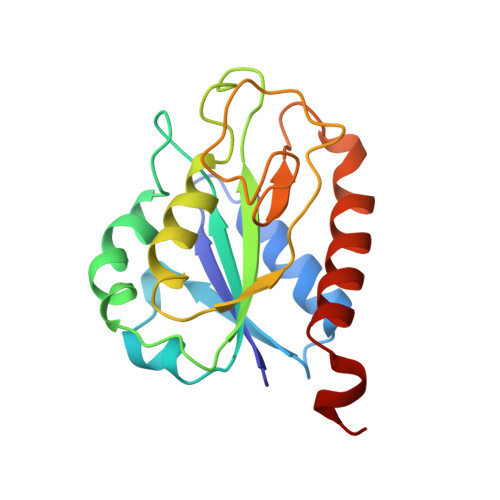
    In Escherichia coli, flavodoxin is the physiological electron donor for the reductive activation of the enzymes pyruvate formate-lyase, anaerobic ribonucleotide reductase, and B12-dependent methionine synthase. As a basis for studies of the interactions of flavodoxin with methionine synthase, crystal structures of orthorhombic and trigonal forms of oxidized recombinant flavodoxin from E. coli have been determined. The orthorhombic form (space group P2(1)2(1)2(1), a = 126.4, b = 41.10, c = 69.15 A, with two molecules per asymmetric unit) was solved initially by molecular replacement at a resolution of 3.0 A, using coordinates from the structure of the flavodoxin from Synechococcus PCC 7942 (Anacystis nidulans). Data extending to 1.8-A resolution were collected at 140 K and the structure was refined to an Rwork of 0.196 and an Rfree of 0.250 for reflections with I > 0. The final model contains 3,224 non-hydrogen atoms per asymmetric unit, including 62 flavin mononucleotide (FMN) atoms, 354 water molecules, four calcium ions, four sodium ions, two chloride ions, and two Bis-Tris buffer molecules. The structure of the protein in the trigonal form (space group P312, a = 78.83, c = 52.07 A) was solved by molecular replacement using the coordinates from the orthorhombic structure, and was refined with all data from 10.0 to 2.6 A (R = 0.191; Rfree = 0.249). The sequence Tyr 58-Tyr 59, in a bend near the FMN, has so far been found only in the flavodoxins from E. coli and Haemophilus influenzae, and may be important in interactions of flavodoxin with its partners in activation reactions. The tyrosine residues in this bend are influenced by intermolecular contacts and adopt different orientations in the two crystal forms. Structural comparisons with flavodoxins from Synechococcus PCC 7942 and Anaebaena PCC 7120 suggest other residues that may also be critical for recognition by methionine synthase.


  • Organizational Affiliation

    Department of Biological Chemistry, University of Michigan, Ann Arbor 48109, USA.


Macromolecules
Find similar proteins by:  (by identity cutoff)  |  3D Structure
Entity ID: 1
MoleculeChains Sequence LengthOrganismDetailsImage
FLAVODOXIN
A, B
175Escherichia coliMutation(s): 0 
Gene Names: FLDA
UniProt
Find proteins for P61949 (Escherichia coli (strain K12))
Explore P61949 
Go to UniProtKB:  P61949
Entity Groups  
Sequence Clusters30% Identity50% Identity70% Identity90% Identity95% Identity100% Identity
UniProt GroupP61949
Sequence Annotations
Expand
  • Reference Sequence
Small Molecules
Ligands 5 Unique
IDChains Name / Formula / InChI Key2D Diagram3D Interactions
FMN
Query on FMN

Download Ideal Coordinates CCD File 
H [auth A],
O [auth B]
FLAVIN MONONUCLEOTIDE
C17 H21 N4 O9 P
FVTCRASFADXXNN-SCRDCRAPSA-N
BTB
Query on BTB

Download Ideal Coordinates CCD File 
I [auth A],
P [auth B]
2-[BIS-(2-HYDROXY-ETHYL)-AMINO]-2-HYDROXYMETHYL-PROPANE-1,3-DIOL
C8 H19 N O5
OWMVSZAMULFTJU-UHFFFAOYSA-N
CA
Query on CA

Download Ideal Coordinates CCD File 
C [auth A],
D [auth A],
J [auth B],
K [auth B]
CALCIUM ION
Ca
BHPQYMZQTOCNFJ-UHFFFAOYSA-N
CL
Query on CL

Download Ideal Coordinates CCD File 
E [auth A],
L [auth B]
CHLORIDE ION
Cl
VEXZGXHMUGYJMC-UHFFFAOYSA-M
NA
Query on NA

Download Ideal Coordinates CCD File 
F [auth A],
G [auth A],
M [auth B],
N [auth B]
SODIUM ION
Na
FKNQFGJONOIPTF-UHFFFAOYSA-N
Binding Affinity Annotations 
IDSourceBinding Affinity
FMN PDBBind:  1AG9 Kd: 1 (nM) from 1 assay(s)
Binding MOAD:  1AG9 Kd: 1 (nM) from 1 assay(s)
Experimental Data & Validation

Experimental Data

  • Method: X-RAY DIFFRACTION
  • Resolution: 1.80 Å
  • R-Value Free: 0.250 
  • R-Value Work: 0.196 
  • R-Value Observed: 0.196 
  • Space Group: P 21 21 21
Unit Cell:
Length ( Å )Angle ( ˚ )
a = 126.4α = 90
b = 41.1β = 90
c = 68.15γ = 90
Software Package:
Software NamePurpose
X-PLORmodel building
X-PLORrefinement
SDMSdata reduction
SDMSdata scaling
X-PLORphasing

Structure Validation

View Full Validation Report


Ligand Structure Quality Assessment 


Entry History 

Deposition Data

Revision History  (Full details and data files)

  • Version 1.0: 1997-12-24
    Type: Initial release
  • Version 1.1: 2008-03-24
    Changes: Version format compliance
  • Version 1.2: 2011-07-13
    Changes: Version format compliance
  • Version 1.3: 2023-08-02
    Changes: Database references, Derived calculations, Other, Refinement description